
This post was earlier cross-posted at Leonid Schneider's site, hence the unfrivolous tone. The version there is improved by Leonid's editing and frame-story.
These curious images first appeared in PubPeer threads four years ago. They are ostensibly Transmission Electron Microscopy enlargements of Ag₂S or Ag nanoparticles, though with the replicated skulls or masks in motion across a backdrop of nanodetritus, they could easily be cover art for a Bauhaus or Wire bootleg album.


The response of the journals' editors, after F.-X. Coudert alerted them to the presence of Hallowe'en-themed 80s prog-rock covers, was mixed. In the case of the left-hand Figure 1c, from Kumari, Chandran and Khan (2014) - the later-submitted of the two, by a few days - the paper was eventually retracted, for this and other repetitions.
 |
| "Self-portrait with Skulls" |
“Author […] stated that they were not duplicated. We accepted his statement and have no information that contradicts it.”They did eventually publish a Corrigendum, replacing Figures 1c and 1d with completely different images. We'll come back to Fig 1d.
The wrong figures were submitted for Fig. 1c and d in the above referenced article. The correct figures are attached below. The authors regret for the error.Did I mention that both journals are published by Elsevier?

Equilibrium and kinetic studies on adsorptive removal of malachite green by the citrate-stabilized magnetite nanoparticles
"Malachite Green" was the hero in a series of Sherlock Holmes-pastiche detective novels from the 1930s. Not many people know that.
Perhaps they are being juggled.

At any rate, the retraction and correction focussed attention on a Scanning Electron Microscope (SEM) image present in both original versions as Figures 1d or 1b, though identified as Ag and as Ag₂S nanoparticles.


And we shouldn't forget "Toxic potential of iron oxide, CdS/Ag₂S composite, CdS and Ag₂S NPs on a fresh water alga Mougeotia sp" (2015), in which Figure 1 is a tour-de-force of versatility. Three of its four panels consist of further enlargements from the same Protean nanoscape, variously rebranded as iron oxide AND CdS AND Ag₂S NP. Did I mention that all these papers have one author in common?

That team's microphotographic virtuosity does not end there. Further anonymous / pseudonymous scrutiny brought to light a sparser selection of nanodetritus in four-fold repetition, variously identified as colloidal-silver nanites; Titania / TiO₂ [Oberon not included]; Ag again; and ZnO. In that fourth appearance the fragments of nanodandruff have been shuffled around and recomposed within the digital image, with particle reduplication to maximize the aesthetic effect.

Our author of interest was not content with the scruffy, slightly-foxed imperfection of these images, and worked with his students in the quest for neatness and technical elegance:
For the past few years my students are working continuously in my laboratory to get hexagonal shaped ZnO NPs and we could achieve the same. The results were so perfect and TEM images were taken until we get very low noise and perfect hexagonal structure. The low noise was avoided by adjusting the contrast of the image.They responded to his challenge with these Platonic visions of geometrical purity:
 |
| "Influence of humic acid on the stability and bacterial toxicity of zinc oxide nanoparticles in water" (2015) |
 |
| "Interaction of colloidal zinc oxide nanoparticles with bovine serum albumin and its adsorption isotherms and kinetics" (2013) |

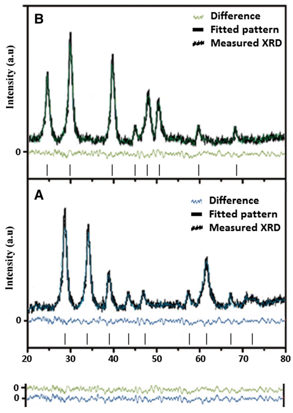
But appreciation of electron-microscopy manipulations should not distract us from the group's real metier, which lies in synthetic spectroscopy. "Unregistered Submission" at PubPeer brought together XRD patterns from five publications (and five materials). Here 'XRD' is X-Ray Diffraction: a crystal reflects an incident beam of X-rays at certain specific angles where the spacing of atoms along some plane within the crystal lattice matches the X-ray wavelength, allowing the crystallographer to characterise that arrangement of atoms and confirm or falsify what it's supposed to be. XRD does not require a single large crystal... like a rainbow, forming from light bouncing off a random cloud of raindrops without the need for a single large prism to refract the sunlight, it can work on a powder of tiny crystals (nanoparticles) in random alignments.

 But there is inevitable noise in any diffractogram: small-scale fluctuations smearing out the flanks of the diffraction-angle peaks and ploughing up the flat baselines between them. Bear in mind that it is the business of
 The earliest XRD trace in this set was from 2013 - at the bottom, here - and it seems to be the original. The material was ZnO. The small peak at 2θ = 18° was surplus to requirements, so in the 2014, 2014 and 2015 versions it was replaced by copy-pasting a small-angle squiggle (conceivably a signature).

Once you taste the joys of copy-pasting squiggles it is hard to stop. Akhil (2016) brought multiple variations on a theme together in one place: ostensibly independent measurements of ZnO NPs, separately synthesized while fine-turing their surfaces with ethylene glycol, polyvinyl alcohol, polyvinylpyrrolidone and gelatin. They differed only in the duplicated stretch spliced into the middle of each exhibit, with the bulk of the 2013 XRD pattern remaining constant.

As crystal-lattice fingerprints, these are nonsense; how could any competent reviewers accept these Photoshop [or MS Paint] confections?

The next two XRD patterns were not reconstructions of the 2013 ZnO original. Both supposedly came from experiments with iron oxide. One is Figure 3(a) from Jagadeesh again; the second is Fig. 1(d) from Mishra, Chandran & Khan (2014) (which is the Malachite Green paper we encountered near the start). Both are Frankenstein-style patchwork productions. Neither is at all authentic.


Finally, a relatively subtle piece of digital retouching. Perhaps there was a gap left by removing a peak in the wrong place, to be bridged with what appear to be small, laterally-compressed Airedales. Closing the circle, this is Figure 1(e) ("XRD pattern of freshly synthesized Ag NPs") from Alam et al. (2014) - the corrected Spectrochimica Acta paper with which we began.

The authors had an opportunity to amend this Figure when they replaced 1(c) and 1(d), but passed it by, so I guess they still stand by 1(e).
As always, I am only showing the shiniest objects from the PubPeer threads, which readers should explore for themselves. More could be said about S. Sudheer Khan's colleagues at the same institution:
This paper is by authors from VIT Vellore. Several papers from this institute in Vellore have been discussed in Pub Peer. In the present case there is an extensive recycing of the same figures / data. It is hoped that the authorities of VIT Vellore will look into this and start taking approriate action to stop this practise.Indeed, the "digital dandruff" image is not original to S.S.K., and its first two appearances [above] were in papers without his co-authorship; his colleagues passed it on when they had finished with it, like a half-sucked lollipop. But anyway, this is really a concern for Indian scientists. I am more interested in the whole saga as a symptom.
* * * * * * * * * * * * * * * * * * * *
Crap papers are written for any number of reasons. The general theme that as long as "science" is seen as a Good Thing, people will be keen to pretend that their mental effluvia count as "science" - even if they would be rejected from collections of Vogon Poetry. So we rely on the journals and their tradition of peer review to function as the Gate-Keepers of Academia... keeping the Literature unsullied by displays of gratuitous incompetence; by word-wooze churned out by seat-warming drones for CV-polishing and career enhancement; by alt-med grifters engaged in cargo-cult science and white-coat cosplay; by fabrications intended to camouflage someone's political agenda as Objective Fact.(thereby forcing all these different kinds of interlopers to launder their productions through journal-shaped dumpsters from predatory publishers, in the form of paid press releases in paper disguise).But here the gate-keeping was an abject failure. The gates are wide open and so many Trojan Horses have trundled into Troy that the streets are congested and there is no space for the townspeople to go about their business. Emblazon the Wall of Shame with the names of the offending journals:
Bioprocess and Biosystems Engineering
Colloids and Surfaces B Biointerfaces (x 4)
Journal of Basic Microbiology (x 2)
Journal of Environmental Sciences
Journal of Photochemistry and Photobiology B Biology (x 5)
RSC Advances
Solar Energy
Spectrochimica Acta Part A
Not the Elsevier logo
A contributing factor could be the dominant market positions of a few publishers. Or it could be the journal editors, who are often fighting to increase the prominence and prestige of their particular specialty (that's why they became editors!), even at the cost of padding out the page-count of their journals with empty pixels. But why bore you with my speculations, when I could be boring you with more examples of research malfeasance?
* * * * * * * * * * * * * * * * * * * *

[updated in proof]
Fakhri's talents include liquid chromatography (Fakhri & Nejad, 2016):
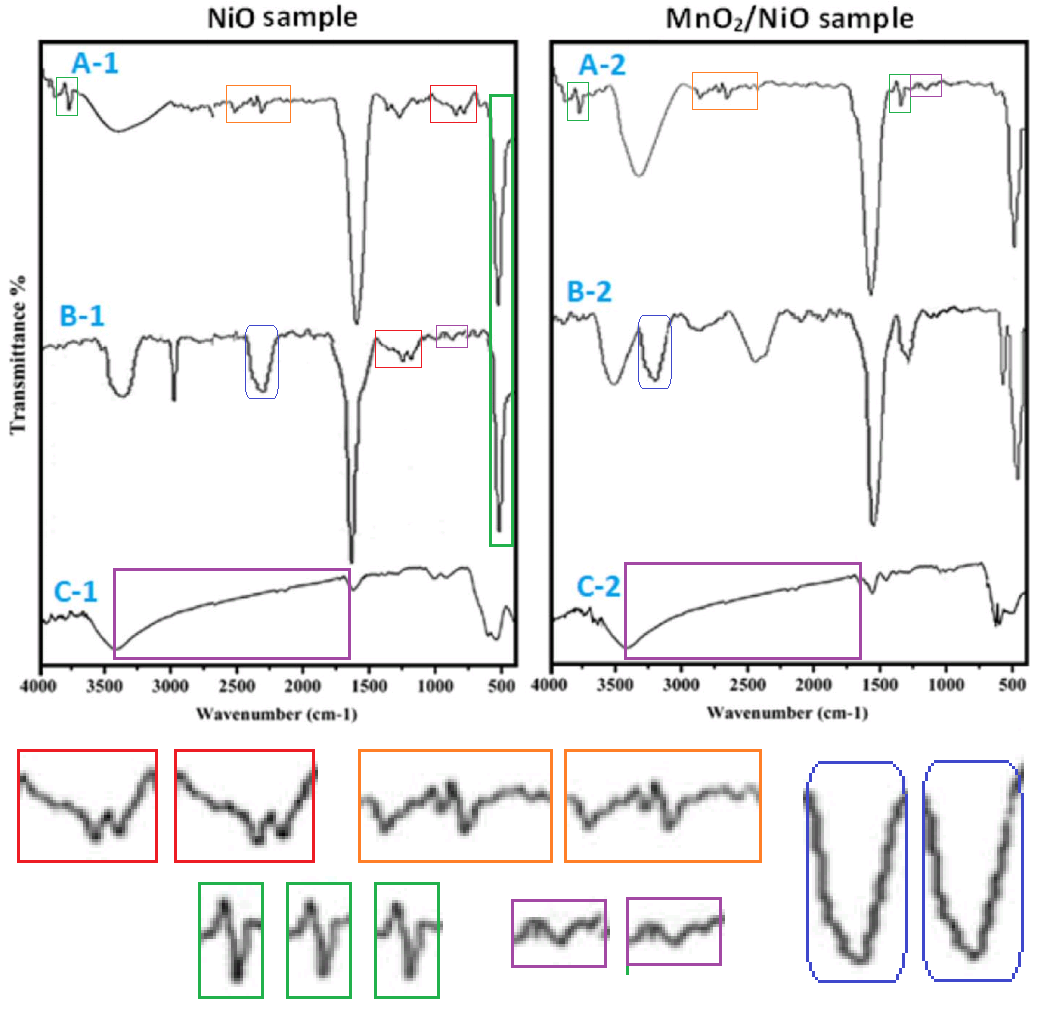
...and patchwork Fourier-Transform Infrared spectra (Gupta et al. 2017)
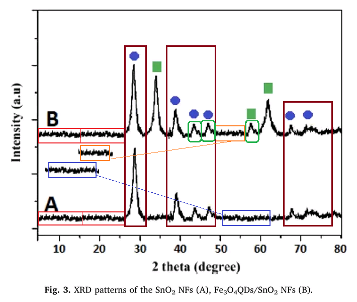
However, his specialty consists of the production of XRD scans for a diversity of nanotechnology materials, plotted in a way that highlights the pixel-level jaggies of noise replicated across them.
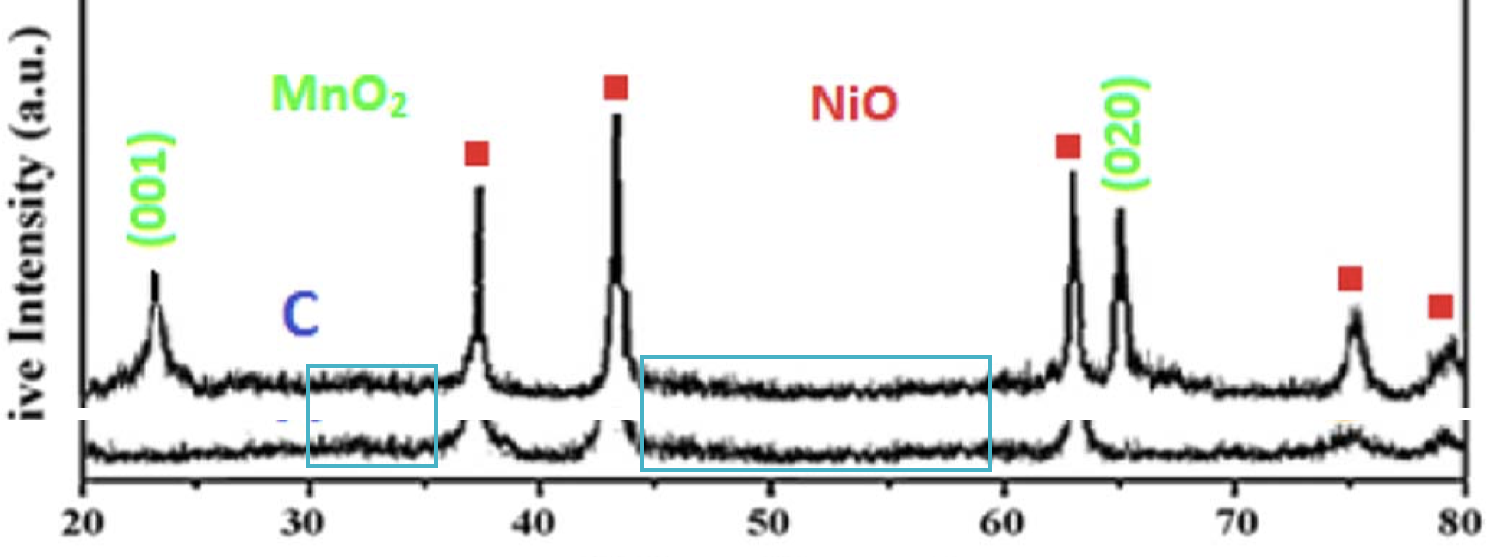
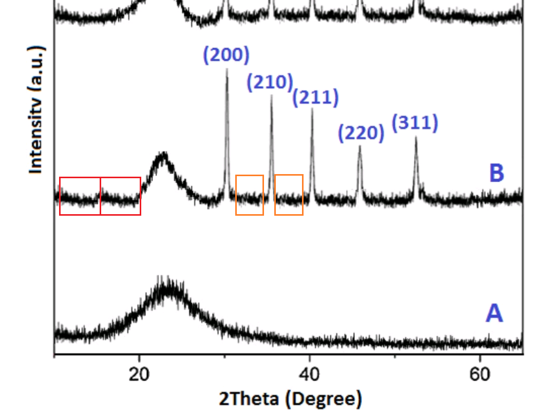
Peaks duplicate and shift location like jumping genes, within single Figures and between papers. They bring to mind the Māori legends of migratory mountains.
 |
| The mountains skipped like rams, and the little hills like lambs. |
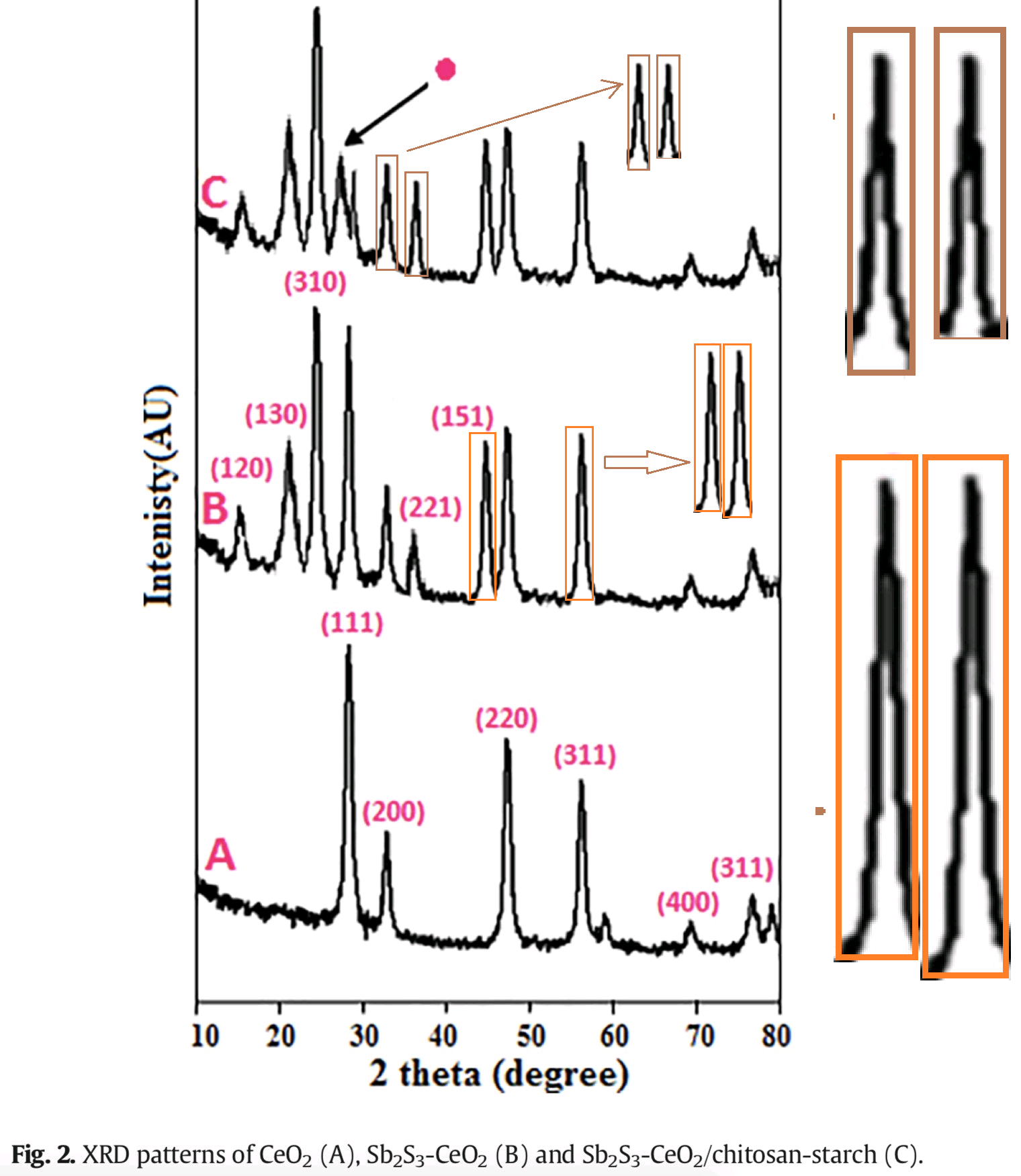
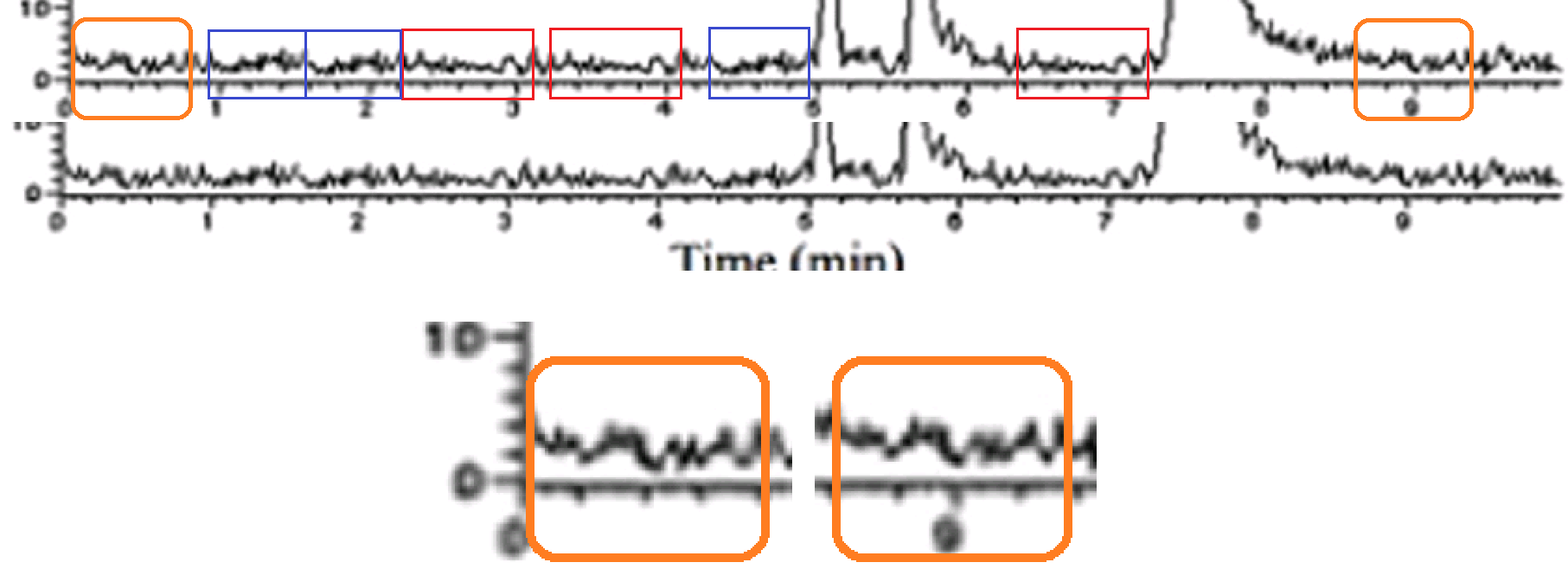
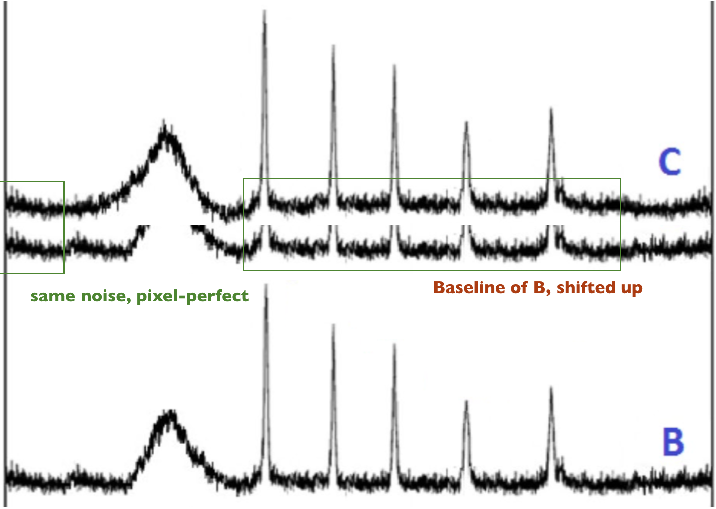
These patterns are especially noteworthy for their baselines - the flat-line, diffraction-free, noise-only stretches between the peaks - where we find the noise repeated like a tape-loop or a music sample in some aspiring rapper's demo track (Fakhri & Kahi 2017).

Two more variations on the theme of graphene-oxide XRD:


Sometimes these reverberant noise sequences are assembled from smaller units, in the manner of trains, and it is tempting to turn them into animated GIFs in which they chug across the diagram trailing smoke. Sometimes they even find their way into XRD patterns made up for more than one paper.


This is what keeps bringing me back to the nanotechnology field (or Advanced Materials Science if you prefer). These are not marginal journals (IJBM has an IF of 3.909). Many of the people publishing in them - perhaps even the majority - are not fakes and frauds. But how are readers to tell which is which?
International Journal of Biological Macromolecules (x 6)
Journal of Colloid and Interface Science (x 2)
Journal of Colloid and Interface Science (x 2)
Journal of Inorganic and Organometallic Polymers and Materials
Journal of Materials Science Materials in Electronics (x 3)
Journal of Molecular Liquids (x 2)
Journal of Molecular Liquids (x 2)
Journal of Photochemistry and Photobiology B Biology (x 9)
Editors and reviewers looked at these 16 papers and thought "Yes, this is how XRD patterns are supposed to appear, full of rap-music samples, migratory mountains and toy train sets". Someone looked at these energy-dispersive Element Maps (Hosseini et al, 2018) and thought "Sulphur atoms in these MnS₂ particles are distributed in multiple copies of Freytag's Pyramid? That seems about right".

Fakhri et al (2019) include "Fig. 3 EDS elemental mapping of the of CoS₂–SiO₂-1/CS nanocomposites":

The Si and O panels, at least, were not obviously constructed from extensive use of the Clone stamp. Mind you, the Cobalt panel mostly looks plausible enough, except for the zone which was filled in with a repeating tow-truck motif.
Having started this post with TEM shenanigans, we should probably finish the same way:


The right-hand image nominally portrays a "CdSe quantum-dots-decorated SnO2 nanotube", but I like to think that it is really a digital plant-stem, suffering from an infestation of Photoshop scale-bugs.

No comments:
Post a Comment