
This post was earlier cross-posted at Leonid Schneider's site, hence the nonfrivolity and Explaining Voice (and the images reduced to nigh-invisibility here). The version there is improved by Leonid's frame story, Tiger Lillies videos, t-shirt design and editing.
 Recent news stories have shown that the way to buttress one's claims with evidence is to copy images and draw over them with a felt-tip pen. In this research tradition, the Milan University group of Desiderio and colleagues were ahead of their time.
So here is a Western Blot electrophoresis image, extant in versions with three, five and four columns, depending on how many lanes have been photocloned and then crudely retouched. They hail respectively from Maroni et al. (2011) (which I assume is the original), Bendinelli et al. (2013) and Maroni et al. (2015) - these are listed below as [6], [14], [9].

Now Desiderio's laboratory group labour in the academic groves of molecular cell biology, and specifically, the subgrove devoted to metastatic breast cancer. They map out the network of metabolic proteins and signalling proteins involved when carcinoma cells go metastatic, or adapt to the hypoxic conditions inside a tumour. Their production of papers displays what one might call a kind of artistic unity, with images reappearing in diverse contexts across a decade, tying that oeuvre together in the manner of The Dude's rug... but also attracting a number of critical threads from the anonymous, incorrigible peanut gallery at Pubpeer.
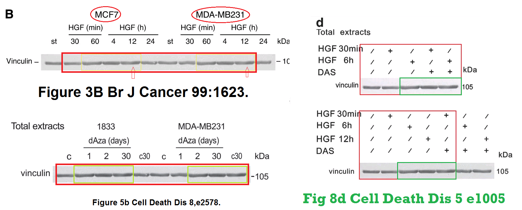
That last sentence is there to foreshadow a "Loading-control Library" theme, but before diving into that, we have not finished with the "retouching" theme. Here's Figure 8(d) from [12] -- Maroni et al. (2014) if you like -- with details enlarged at the right:


The query from "Heterorhabditis Downes" on August 20 evoked a brief rebuttal from the last author on September 2:
The bands are similar but they are not the same.Turning to Bendinelli et al. (2011) [7], the modifications to Figure 3(b) are more subtle, hinting of the airbrush more than the felt-pen. They might go unnoticed without contrast enhancement.

But all of Fig 3(b) deserves to be seen -- not just the single panel shown (twice) above. In the unavoidably over-sized diagram below, Fig 3(b) is right of centre. Left of centre is Fig 4A from Matteucci et al. (2013a) [10]; at top, Fig 2A from Bendinelli et al. (2014) [13]; and at bottom, Fig 9A [Hypoxia] from Maroni et al. (2011) [8]. Pay special attention to the 'vinculin' bands, which are the loading controls -- ensuring equal cell-lysate concentrations across the lanes -- made memorable in this case by the repeating 'sea-serpent' motif. The Desiderio group prefer vinculin for this purpose on account of its predictable good behaviour.
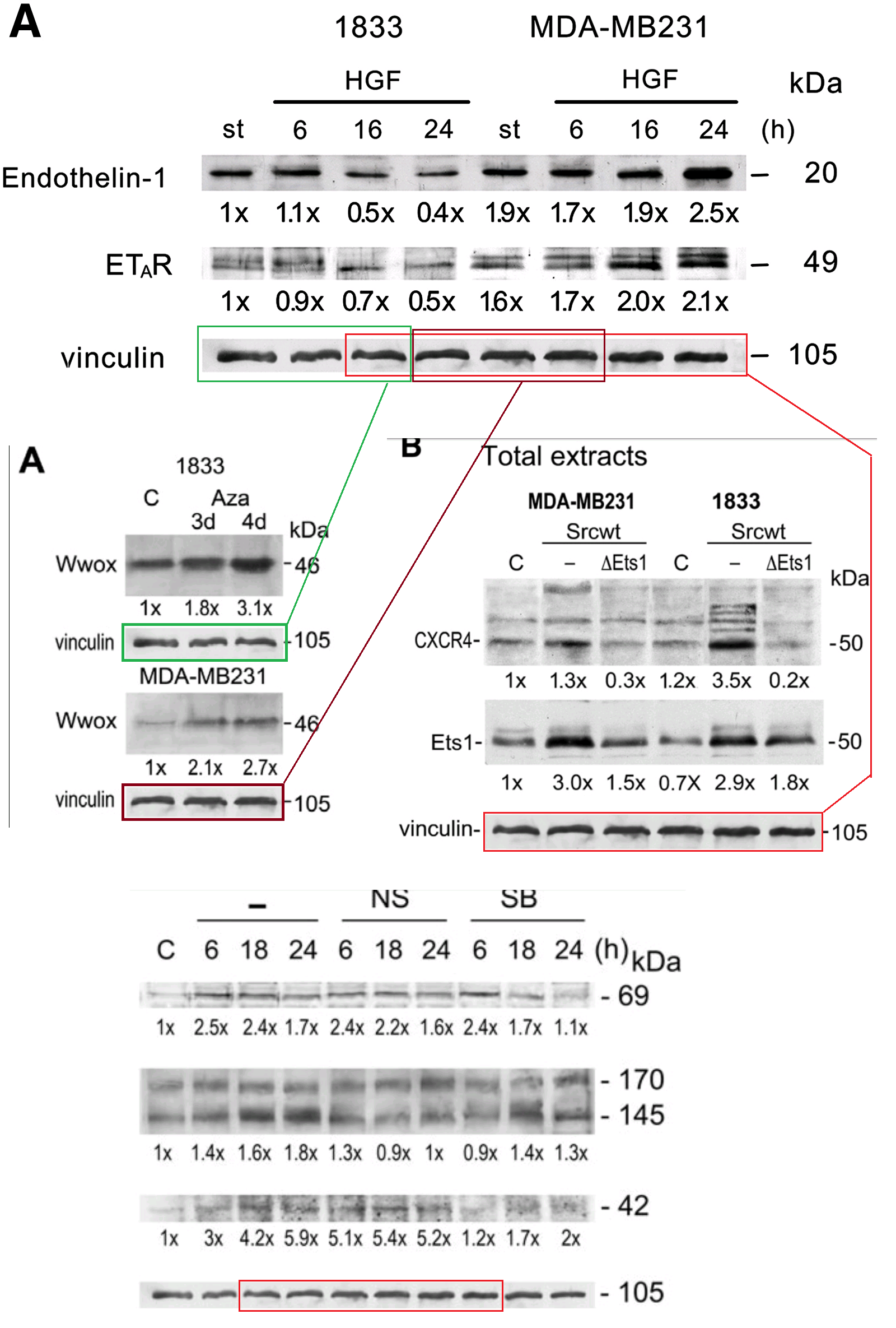
Evidently I am not alone in thinking "Ave Vinculin Invictus!" every time that useful, versatile protein resurfaces in a new context.

But we are not finished with [7]. 'Peer 1' wondered about the similarity in Figure 4D, between the 7th / 8th lanes of the 'vinculin' band and the 3rd / 4th lanes.

The same band had appeared slightly earlier in Previdi et al. (2010) [5], not endowed with duplicated lanes because that experiment only had six conditions for it to ensure equal loading across. See below, left. While below right, more airbrushing hides the join in Fig 4(b), between two copies of a single lane (serving to illustrate different treatments).


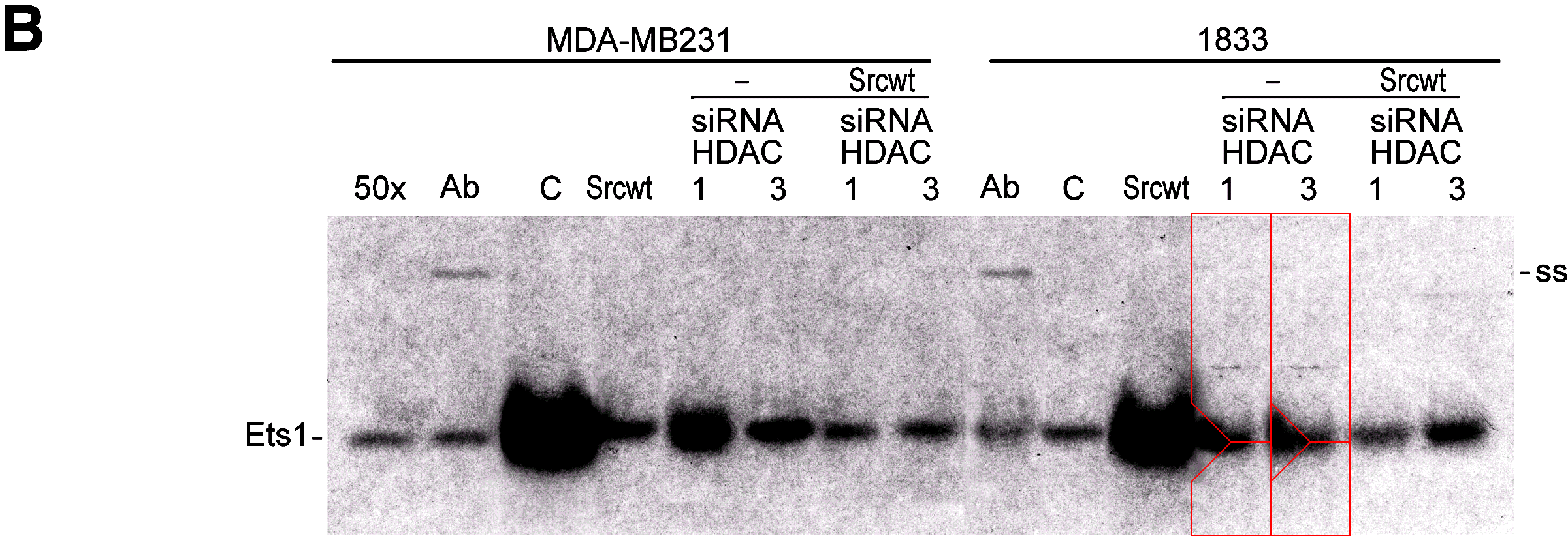
Peer 1's concerns with [7] reached the authors who sought to assuage them, not seeing any serious issue:
Figure 4B BBA 1813: We do not understand why the bands in the squares are evidenced because they are insignificant.Subsequent questions went unanswered, to the disappointment of anyone who hoped to learn the authors' current interpretation of Figure 1A. There, cultured carcinoma cells were enticed to migrate through a nutrient gel in a "Matrigel invasion assay", to test the migration-deterrent properties of different treatments:
Figure 4D BBA 1813: It looks like a technical problem of the photographer in the preparation of the Figure also because there is a number “1” enclosed without significance.


Thus we encounter another of the unifying themes in the Desiderio oeuvre. Like a single small fish in a bottle of milk, the presence of a single duplicated cell in a Matrigel invasion assay is enough to erode one's faith in the veridical nature of the composition, and often there is more than one. From Matteucci et al. (2013b) [11], part of Fig 4E:
Figures 3B, 3D:


Also from 2013, here are the two right-hand panels from Fig 4E of [10]. The two left-hand panels can be omitted here as they duplicate Fig 3D from [11] (albeit rotated through 90° and labeled with different cell counts). We can also omit Fig 2D as its panels reappeared in 2015 as Fig 1C of [14].

Moving on to 2014, and [13], lovingly-curated collations of cells feature in Figures 1D, 2B, and 3A -- someone in the laboratory really enjoys composing them. Cells are spaced at implausibly-regular intervals, copied within but also between panels, increasing the resemblance to wallpaper. We only have space to admire 2B.
In contrast, the panels of Fig 1C from [14] remind me more of leopard-pattern faux-fur. If I were a student at the University of Milan, I would screen-print some of these collages onto t-shirts and sell them from a stall outside the Department of Biomedical Sciences for Health.
This whole line of enquiry was triggered by 'Actinopolyspora Biskrensis' when he scanned Fig 3D from Matteucci et al. (2018) [19] with the 'Forensically' duplicate-detection tool.
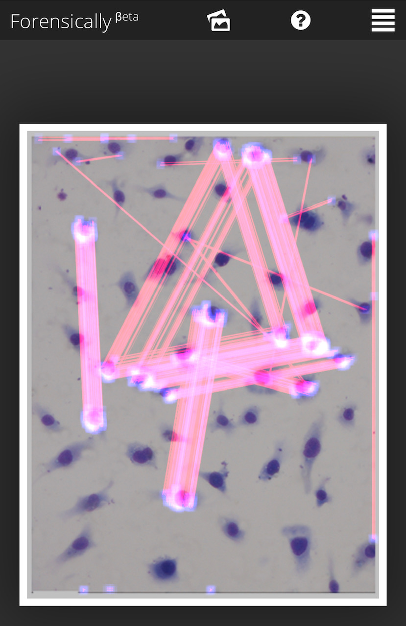
Evidently Forensically only reported photocloning within one single panel of Fig 3D. This could be taken as a challenge to see if the


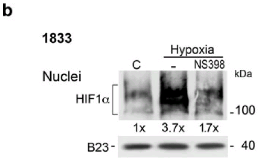
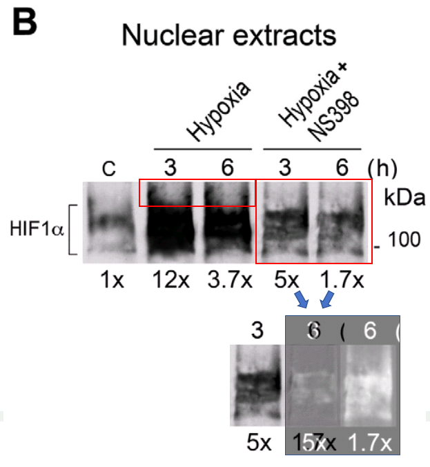
With the 'glitch in the Matrigel' theme finally out of the way (there are Invasion-assay images from 2008 and 2009 but the resolution is too low to make them worth scrutinising), we can return at last to the "Loading-control Library" theme. The Milan team accept that a loading control is a desirable adornment to an electrophoresis result, removing accidental or unconscious differences between experimental conditions and reassuring the reader that the researchers have not led themselves astray. But they are reluctant to waste time and laboratory resources creating them (vinculin might be a bugger to work with, for all I know), relying instead on a small library of equally-loaded bands, cutting off the desired length as if buying six or eight or 10 links of sausage from a butcher.
Did someone say "links of sausage"?
(see Fig 2 of [15])
(Fig 8C of [2]; then 3B; Fig 5B of [18]; Fig 8 of [12])

(Fig 4A of Matteucci, Bendinelli & Desiderio (2009) [4]; Fig 4B of [10]; Fig 3B of [13])
Fans of "Struwwelpeter" can think of them as collections of severed thumbs if they want an alternative metaphor.
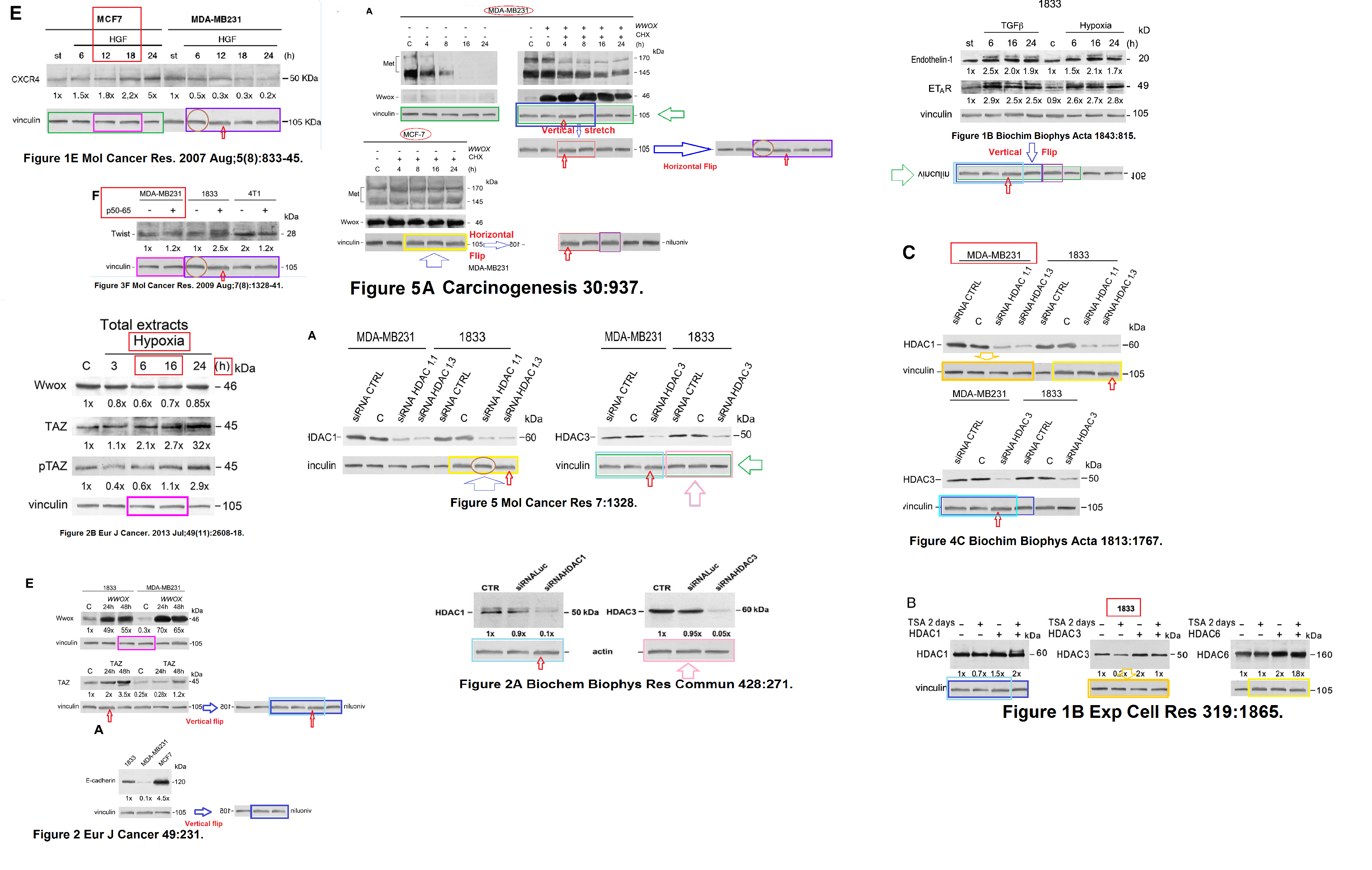
We have already met the 'sea-serpent' motif, so now here is another oversized diagram, bringing together manifestations of a vinculin band most closely resembling a trail of burnt matches. The sightings span almost a decade -- from Ridolfi et al. (2008) [2], [4], through Bendinelli et al. (2009) [3], [12], Bendinelli et al. (2015) [15], to Matteucci et al. (2016) [17] and most recently Bendinelli et al. (2017) [18].

The band is flipped horizontally in one case, and often chopped into smaller sections as if visited by "The great, long, red-legged scissorman" and then reassembled (depending on the number of experimental lanes requiring calibration), but the shapes of the burnt matches are distinctive.
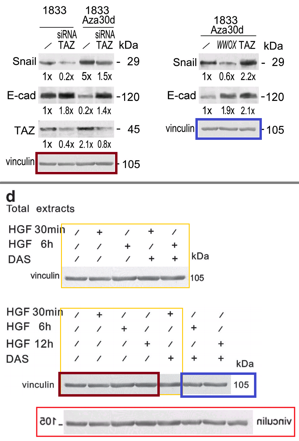
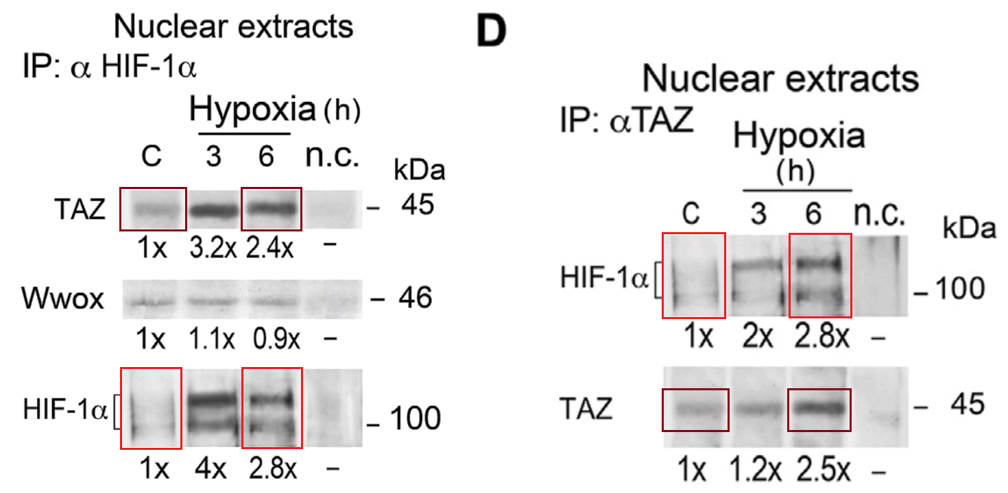
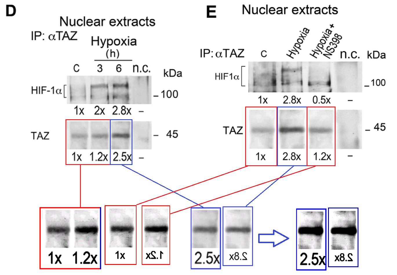
This is all leading up to the mother of all unavoidably oversized diagrams, in which 10 papers shared fragmentary glimpses of yet another library band. The creator (Clare Frances?) described it as "A slightly tangled web".
The authors accepted that the loading bands were erroneous but made no promise to correct them ¯\_(ツ)_/¯. For the burnt-match manifestations:
It looks like an inattention in the graphical assembling of the acquisitions, since the aspect of the vinculins is very similar. In any case the data are calculated on a triplicate.An uncredited student was to blame for the Tangled Web.
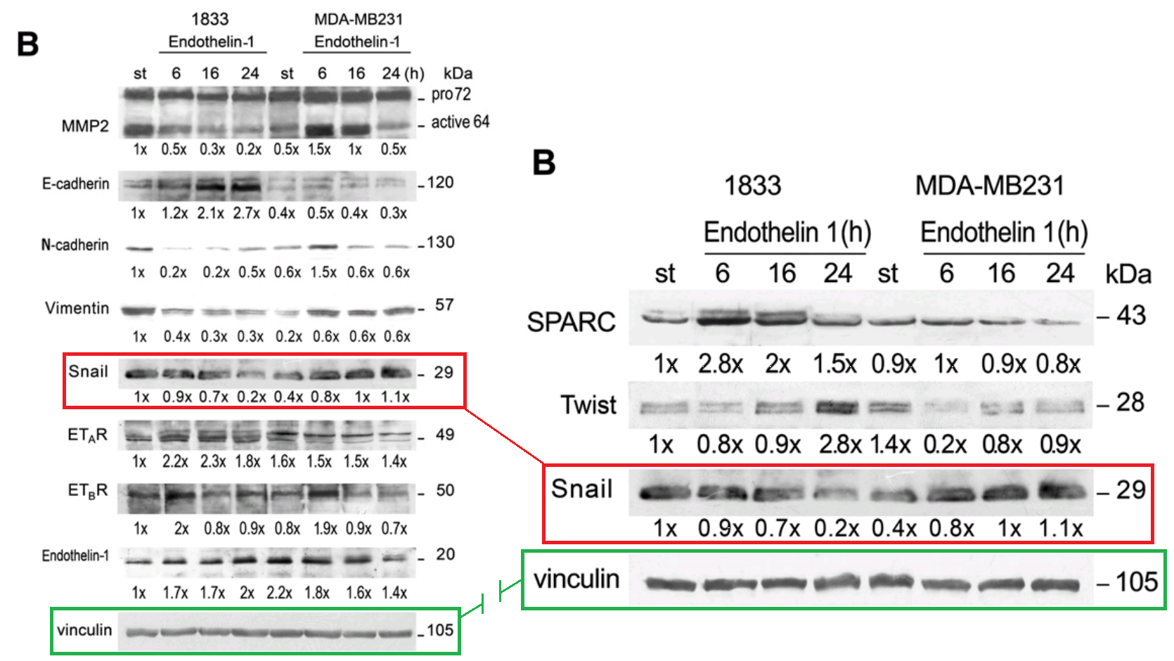
A Phd who does not work any more in the group prepared these Figures.Just saying, there would be no need to "assemble the acquisitions" if they hadn't been cut into small pieces. Things got so confusing that two different loading bands were used in Fig 3B of [13] and 7B of [17] -- sausages / severed thumbs in the former case -- to control for expression of the same Snail band.
Although the recurring themes within this corpus of work all have their illustrations now, I am unwilling to stop, for I do not know if I have conveyed its curious timelessness. The repurposing and re-splicing and retouching of images span the decade. One can begin anywhere in the network and finish anywhere by following the threads between Figures.
Begin, by way of example, with Matteucci, Bendinelli & Desiderio (2009) [4]. We pause to admire its most notable feature, Fig 2A -- an imposing palisade of immunoprecipitated protein fragments. Close, contrast-enhanced inspection reveals the internal repetitions and symmetries that betray its origins in Photoshop.

(Credit: Pardosa Agrestis and Leucanella Acutissima)
But do not let this distract us from Fig 5A, a short train of Wwox blobs. Later the train lost its three central carriages to become part of Fig 1B in Maroni et al. (2015) [14], with the train and caboose directly conjoined (in 2013, 1B was also Fig 2E in [10]).

Within [9], Fig 2D invites further comparison with Fig 4E. Lanes have been copied, flipped horizontally, and darkened as if by police constructing an identity line-up.

 In keeping with this nascent tradition, Fig 2C (still at [9] by the way) resembles an identity line-up of suspension-bridge towers... "Do you recognise any of these as the Brooklyn Bridge you bought?" But few readers will be surprised to discover that Lane 7 is a horizontally-flipped, retouched version of Lane 4.
Then [9] leads back to [4], by way of a reused B23 loading control for Western blots of nucleus proteins,
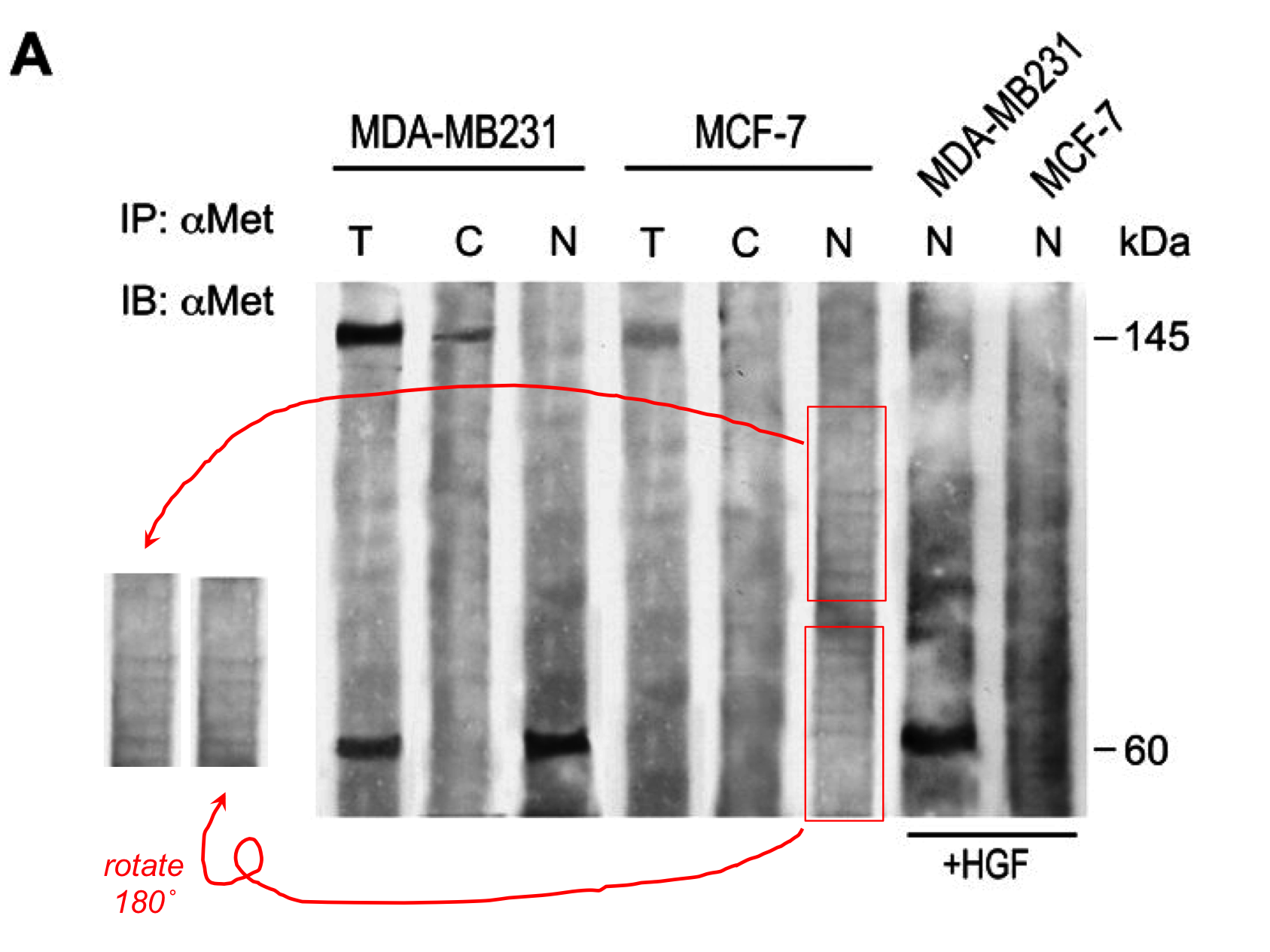
 and a HDM2 band that appears to have been assembled from pieces of a TAZ band (flipping one piece and rotating another through 180°). But I don't have time to explain that because the Editor is waiting impatiently for this post. Nor is there time to explain the diagram below right, where [4] jumps back a decade, by way of Fig 1D and the lanes it shares with Fig 4B from Maroni et al. (2006) [1].
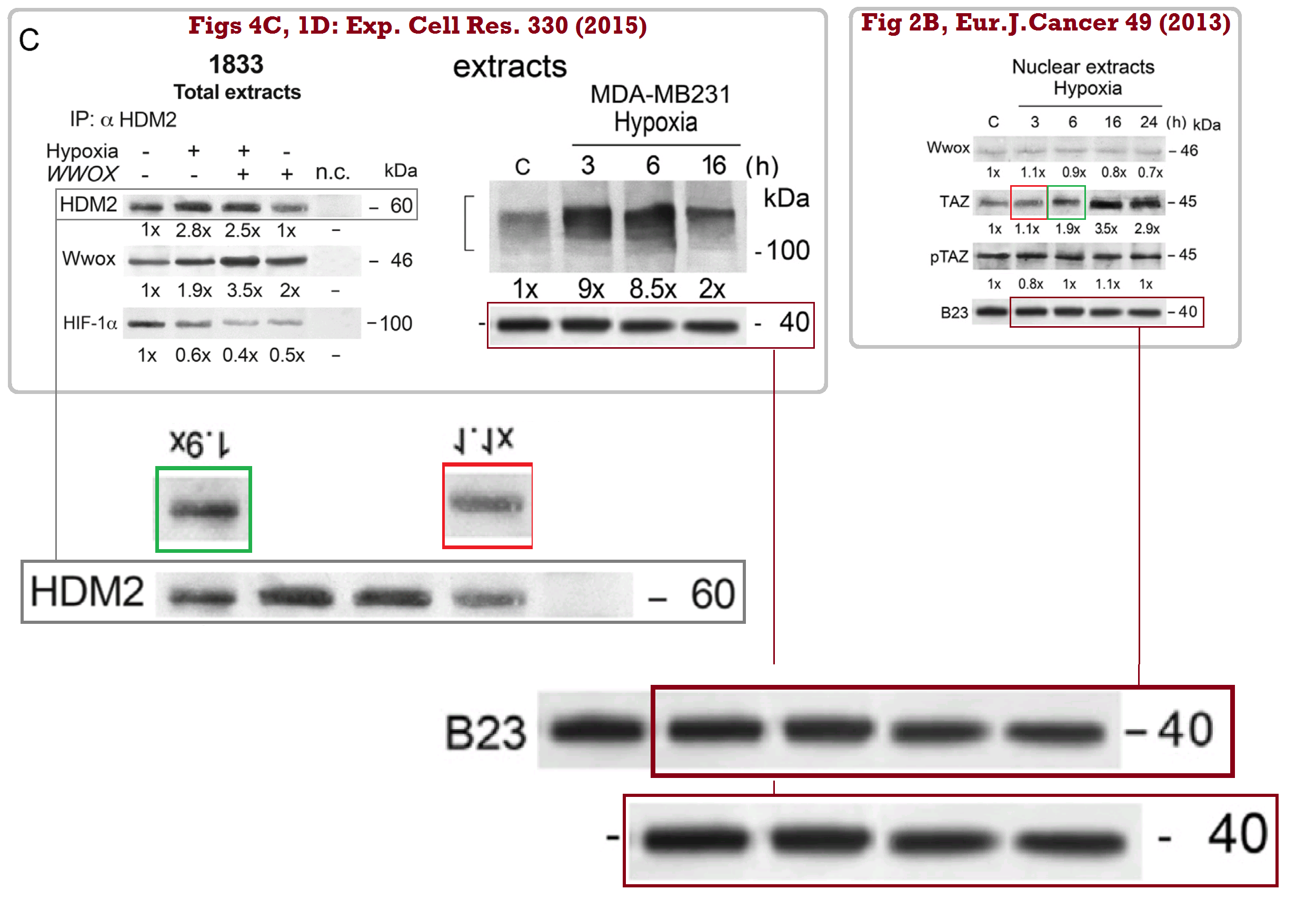

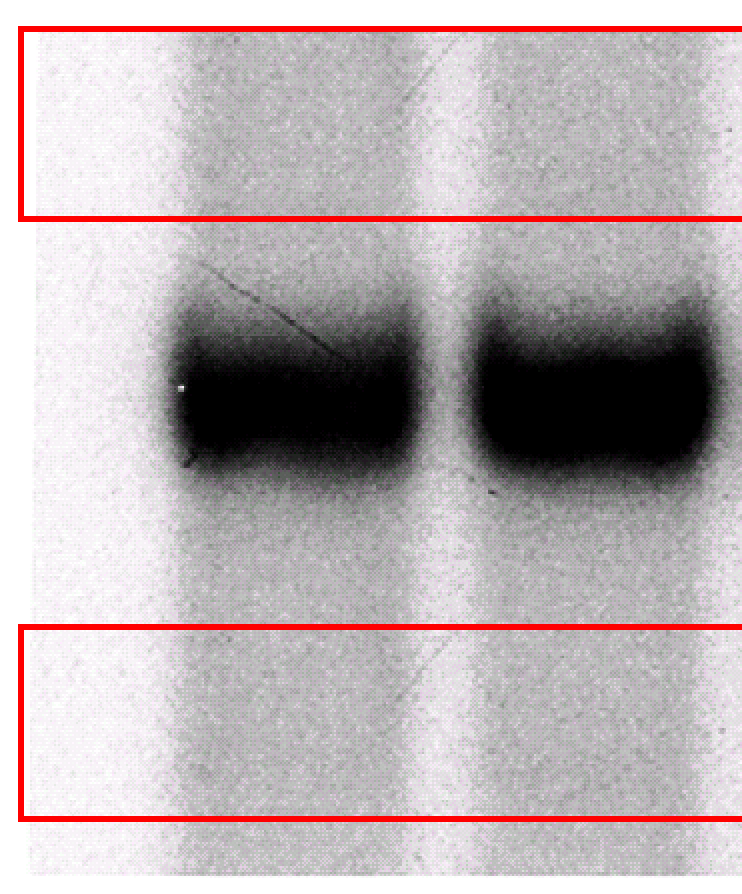
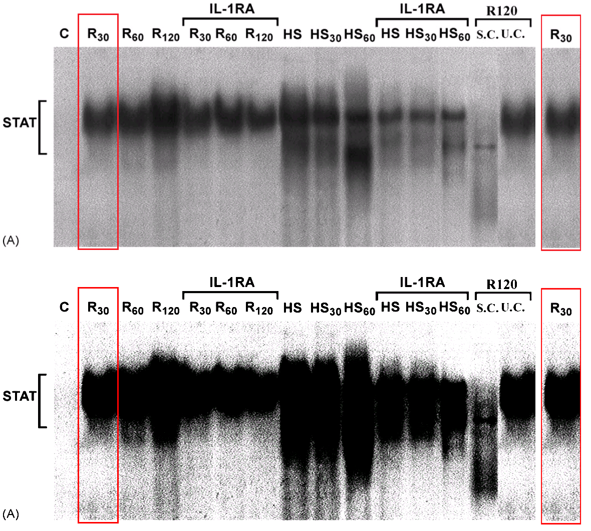
[1] in turn leads back to [3] from 2009, through forensic dental X-ray comparisons of their RNA blots.

For a change of pace, how about some mice, from [10]?
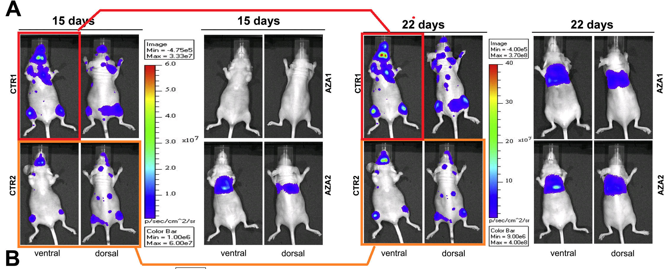
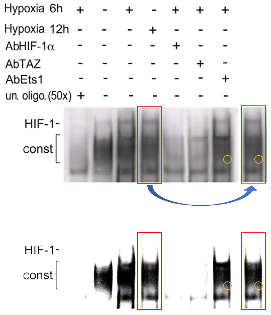
"Am I missing something with Figure 6A? It appears that for mouse Control 2, the Dorsal and Ventral views are the same at 15 and 22 days, although greater bioluminescence is shown at 22 days. For Control 1, the Dorsal views are different but the Ventral views are the same at 15 and 22 days... but again, greater bioluminescence is claimed for 22."I have been neglecting [19], the most recent in this series. In Fig 1E, "Is there some reason why the upper and lower sections of each lane are identical?" In Fig 5B, "the upper and lower extensions of each lane display identical fine texture. Is this a common phenomenon?"


I didn't say they were good excuses.
Sources:
[1]. "HGF induces CXCR4 and CXCL12-mediated tumor invasion through Ets1 and NF-kappaB", Paola Maroni , Paola Bendinelli , Emanuela Matteucci , Maria Alfonsina Desiderio (2006).
Carcinogenesis doi: 10.1093/carcin/bgl129 [PubPeer]
[2]. "Inhibitory effect of HGF on invasiveness of aggressive MDA-MB231 breast carcinoma cells, and role of HDACs", E Ridolfi, E Matteucci, P Maroni, M A Desiderio (2008).
British Journal of Cancer doi: 10.1038/sj.bjc.6604726 [PubPeer]
[3]. "NF-kappaB activation, dependent on acetylation/deacetylation, contributes to HIF-1 activity and migration of bone metastatic breast carcinoma cells", Paola Bendinelli , Emanuela Matteucci , Paola Maroni , Maria Alfonsina Desiderio (2009).
Molecular Cancer Research doi: 10.1158/1541-7786.mcr-08-0548 [PubPeer]
[4]. "Nuclear localization of active HGF receptor Met in aggressive MDA-MB231 breast carcinoma cells", Emanuela Matteucci , Paola Bendinelli , Maria Alfonsina Desiderio (2009).
Carcinogenesis doi: 10.1093/carcin/bgp080 [PubPeer]
[5]. "Interaction between human-breast cancer metastasis and bone microenvironment through activated hepatocyte growth factor/Met and beta-catenin/Wnt pathways", Sara Previdi , Paola Maroni , Emanuela Matteucci , Massimo Broggini , Paola Bendinelli , Maria Alfonsina Desiderio (2010).
European Journal of Cancer doi: 10.1016/j.ejca.2010.02.036 [PubPeer]
[6]. "Nuclear co-localization and functional interaction of COX-2 and HIF-1α characterize bone metastasis of human breast carcinoma". Paola Maroni , Emanuela Matteucci , Alessandro Luzzati , Giuseppe Perrucchini , Paola Bendinelli , Maria Alfonsina Desiderio (2011).
Breast Cancer Research & Treatment doi: 10.1007/s10549-010-1240-1 [PubPeer].
[7]. "Comparative role of acetylation along c-SRC/ETS1 signaling pathway in bone metastatic and invasive mammary cell phenotypes", Paola Bendinelli , Paola Maroni , Emanuela Matteucci , Maria Alfonsina Desiderio (2011).
Biochimica et Biophysica Acta doi: 10.1016/j.bbamcr.2011.06.004 [PubPeer]
[8]. "Nuclear co-localization and functional interaction of COX-2 and HIF-1α characterize bone metastasis of human breast carcinoma", Paola Maroni, Emanuela Matteucci, Alessandro Luzzati, Giuseppe Perrucchini, Paola Bendinelli, Maria Alfonsina Desiderio (2011).
Breast Cancer Research & Treatment doi: 10.1007/s10549-010-1240-1 [PubPeer]
European Journal of Cancer doi: 10.1016/j.ejca.2013.03.002 [PubPeer].
[10]. "Bone metastatic process of breast cancer involves methylation state affecting E-cadherin expression through TAZ and WWOX nuclear effectors", Emanuela Matteucci, Paola Maroni, Alessandro Luzzati, Giuseppe Perrucchini, Paola Bendinelli, Maria Alfonsina Desiderio (2013a).
European Journal of Cancer doi: 10.1016/j.ejca.2012.05.006 [PubPeer]
[11]. "Epigenetic control of endothelin-1 axis affects invasiveness of breast carcinoma cells with bone tropism", Emanuela Matteucci , Paola Maroni , Paola Bendinelli , Alessia Locatelli , Maria Alfonsina Desiderio (2013b).
Experimental Cell Research doi: 10.1016/j.yexcr.2013.04.022 [PubPeer]
Cell Death & Disease doi: 10.1038/cddis.2013.465 [PubPeer]
[13]. "Microenvironmental stimuli affect Endothelin-1 signaling responsible for invasiveness and osteomimicry of bone metastasis from breast cancer", Paola Bendinelli, Paola Maroni, Emanuela Matteucci, Alessandro Luzzati, Giuseppe Perrucchini, Maria Alfonsina Desiderio (2014).
Biochimica et Biophysica Acta (BBA) - Molecular Cell Research [PubPeer]
[14]. "Hypoxia induced E-cadherin involving regulators of Hippo pathway due to HIF-1α stabilization/nuclear translocation in bone metastasis from breast carcinoma", Paola Maroni , Emanuela Matteucci , Lorenzo Drago , Giuseppe Banfi , Paola Bendinelli , Maria Alfonsina Desiderio (2015).
Experimental Cell Research doi: 10.1016/j.yexcr.2014.10.004 [PubPeer].
[15]. "HGF and TGFβ1 differently influenced Wwox regulatory function on Twist program for mesenchymal-epithelial transition in bone metastatic versus parental breast carcinoma cells",
Paola Bendinelli, Paola Maroni, Emanuela Matteucci, Maria Alfonsina Desiderio (2015).
Molecular Cancer doi: 10.1186/s12943-015-0389-y [PubPeer]
[16]. "The Autophagic Process Occurs in Human Bone Metastasis and Implicates Molecular Mechanisms Differently Affected by Rab5a in the Early and Late Stages", Paola Maroni, Paola Bendinelli, Massimo Resnati, Emanuela Matteucci, Enrico Milan, Maria Alfonsina Desiderio (2016).
International Journal of Molecular Sciences doi: 10.3390/ijms17040443 [PubPeer]
[17]. "Coordinate regulation of microenvironmental stimuli and role of methylation in bone metastasis from breast carcinoma", Emanuela Matteucci , Paola Maroni , Andrea Disanza , Paola Bendinelli , Maria Alfonsina Desiderio (2016).
Biochimica et Biophysica Acta doi: 10.1016/j.bbamcr.2015.10.010 [PubPeer]
Cell death & disease doi:10.1038/cddis.2016.403 [PubPeer]
International Journal of Molecular Sciences doi: 10.3390/ijms19010258 [PubPeer]
----------------------------------------------------------
Comment / Update #1:
Here’s yet another Desiderio Library Band, working its loading-control magic in six papers from 2008 to 2013:

Comment / Update #2:
However, in this case the comment of Pandanus Comatus about 18S RNA seems inappropriate because it regards two independent papers with no authors in common. I cannot see how the original image could have been duplicated. This casts some doubts about the accuracy of software-based image comparisons.”Prof Cairo is indignant (or at least not entirely dignant) at the suggestion that Fig 2C in a 2004 paper by Tacchini and Desiderio could be in any way connected to Fig 3c in his own 2006 paper. His disavowal of Tacchini, and the argument that the resemblance must be merely a coincidence, are both weakened by the existence of a second parallelism between Figures 2A and 3b respectively.
Comment / Update #3:
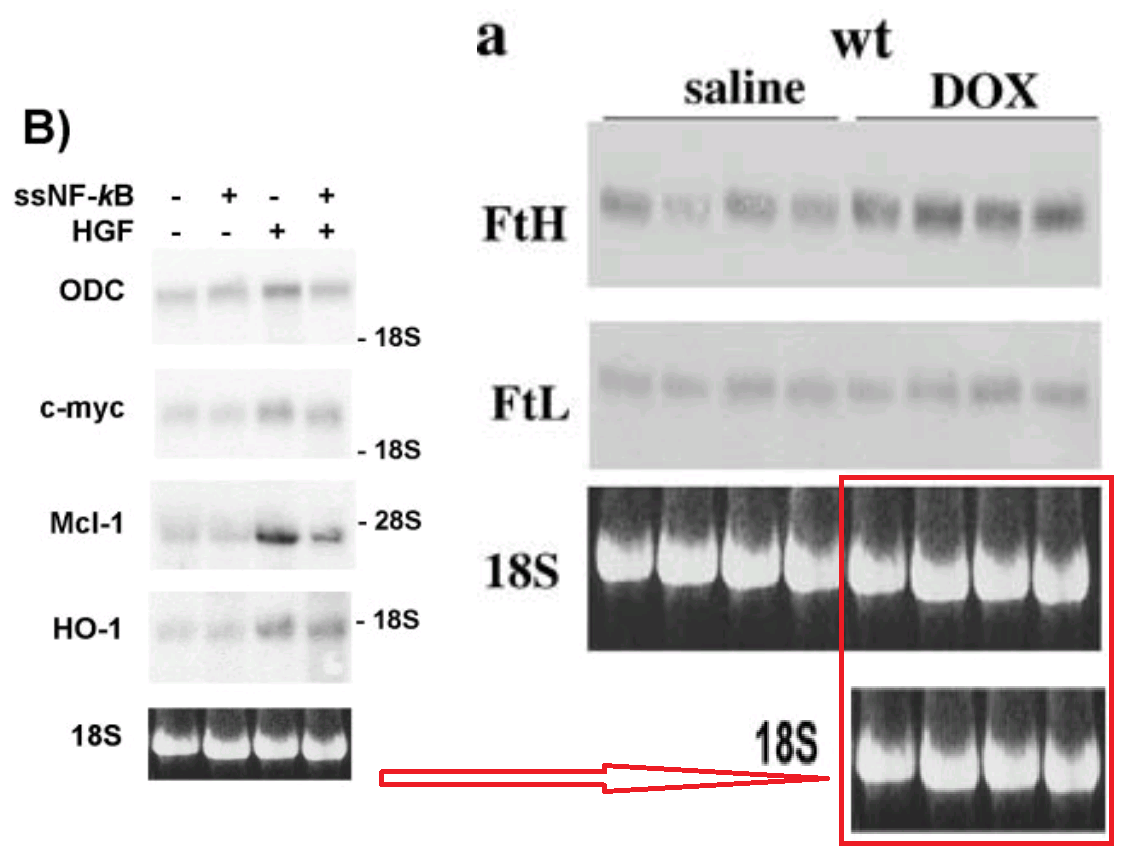
[1] in turn leads back to [3] from 2009, through forensic dental X-ray comparisons of their RNA blots.
My favourite part of the Dental-X-ray RNA blots was a comment from Prof Desiderio, explaining that they were only for purposes of illustration… artists’ conceptions, as it were. She seemed to be genuinely puzzled that anyone could expect them to be factual.
Figure 2A Mol Cancer Res 7 and Carcinogenesis 28: The RNA 18S and 28S in the yellow square were only illustrative of RNA visualization.The texture in the dark background of these blots (visible with increased lightness) is so similar from one lane to another, that they’re quite possibly all constructed by photocloning just one lane...
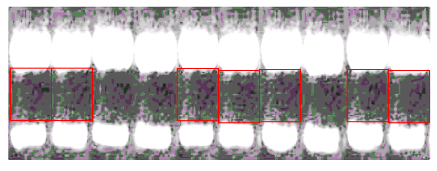
... and then retouching the detailed outlines of the “teeth”.

Comment / Update #4:
Bernelli Zazzera sought to extend his reach over public and charity money by recruiting the bigwig Alberto Mantovani to the University of Milan. Here is an example of the joint output, Tacchini et al 2003
That particular publication has other surprises:
No comments:
Post a Comment