
This post was earlier cross-posted at Leonid Schneider's site, hence the nonfrivolity and Explaining Voice. The version there is improved by Leonid's editing, framing story and explanation of the back-story.
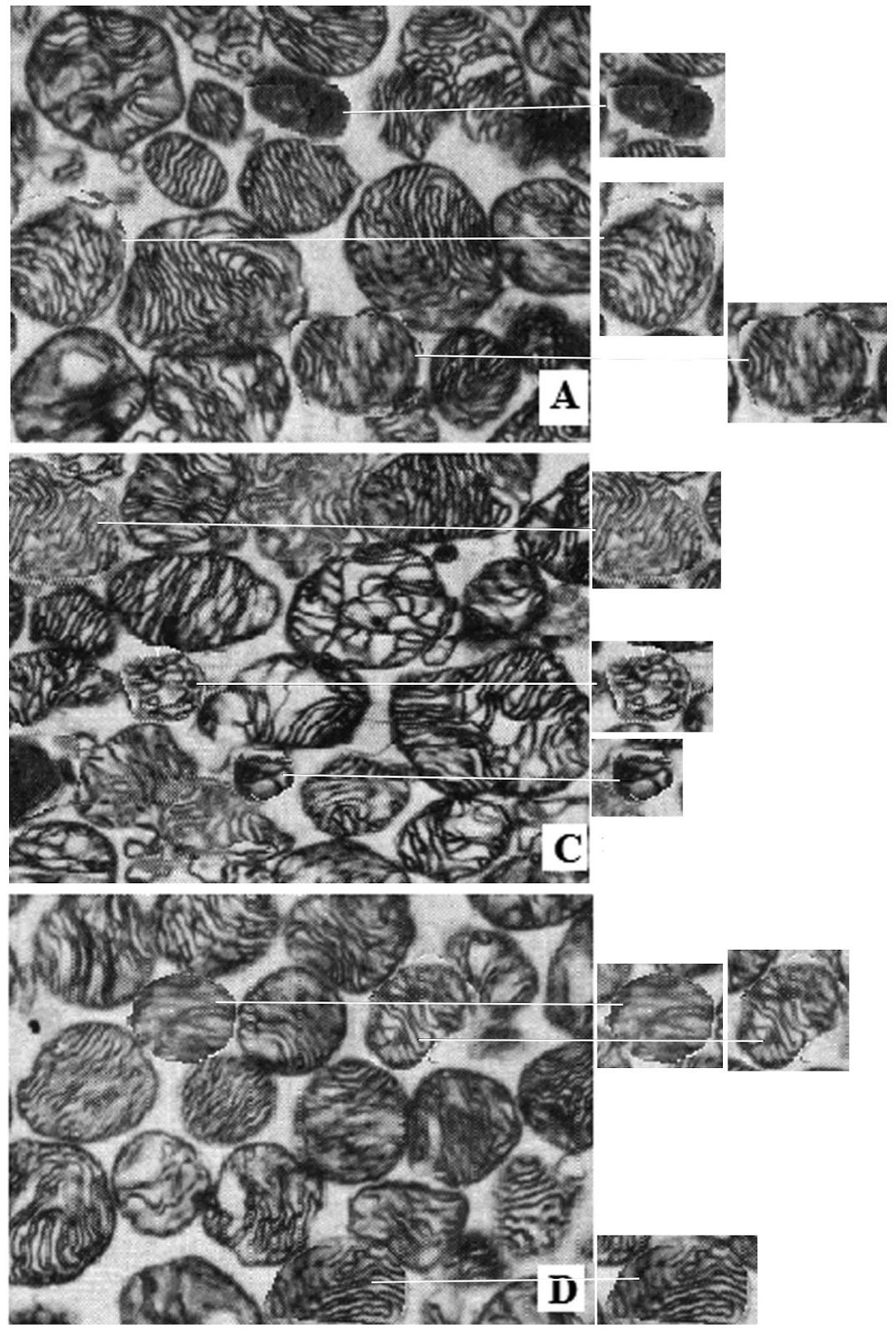
It is hard to look away from these Transmission Electron Microscope images. I am half-convinced that they are really high-contrast Gestalt-perception puzzles and that if I stare long enough they will resolve into a Dalmatian trotting across a dappled background. They are either hippocampal neurons or glomerular podocytes, subjected to oxidative stress by either fluorine or arsenic, ameliorated (or not) by either EGCG (Epigallocatechin gallate) or Sulforaphane (Thangapandiyan et al, 2018 [15]; Thangapandiyan et al. 2019 [16]).


The blotchy markings on two nuclei are in fact the map for a fantasy novel set on a mainly-ocean archipelago planet.

Closer attention reveals more graphic elements recurring within and between the images, and quasi-symmetries within them, arousing the melancholic suspicion that the cells never existed at all outside Photoshop.

 The authors alluded in comments in PubPeer threads to the external provenance of data, explaining that they lack sophisticated software for detecting duplications and fabrications in the material provided by those sources.
#4 S. Miltonprabu
...Repetitions that you have portrayed in the figures were not created by us ... Regarding histopathological studies, the tissues were fixed and despatched the out sourcing lab for prepared the slides and a pathologists would take the photograph from different groups and send their results with comments along the slides. ... Even without any proper sophisticated instruments, with limited funding opportunities, we have done lot of very good works based on the out sourcing process only.
#8 S. Miltonprabu
we are not having the sophisticated software's to identify the images taken from the same slides.Even if it occurred like this, we will replace it with our original slides,if it is requested from the journal, because now we have equipped with our own photo microscopy, we didn't depend on the out sourcing fellows to take the photography our slides.
Professor Miltonprabu is now at Madras University though he was previously at Annamalei University (Kerala State, India) where the present work was conducted, and some of his papers feature in Elisabeth Bik's overview of the research culture at that institution. He lists several research programs on his ResearchGate profile, all following a generic format:
- An oxidative-stressful toxin (arsenic, cadmium, fluoride);
- A vulnerable tissue or cell class (liver, lungs, kidney, cardiomyocytes); and
- A protective botanical extract (grape-seed proanthocyanidins, epigallocatechin gallate, milk thistle, quercetin, diallyl trisulfide from garlic, sulforaphane from the crucifer / cabbage family...)


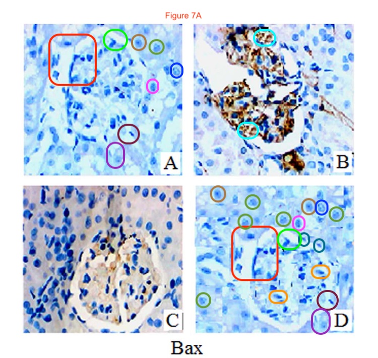
The value of the nuclei in panels (A) and (D) as maps or globes for fantasy planets is reduced by excessive rotational symmetry. Other large chunks repeat between panels with 90° rotation, while Dr Bik's attempt to mark up all the smaller repeated elements have peppered the Figure with so many rounded-off color-coded rectangles that at first I mistook it for an exercise in Egyptology and I wasted a lot of time trying to read the hieroglyphic Pharaoh's name within each cartouche.
Also from [14], Figs 8A and especially 7A could be enlargements from a Pointilliste canvas by Seurat or Signac, with their blue palette and their regular broad brushstrokes. Perhaps too regular. They would make good characters for a decorated-alphabet font consisting entirely of 'O's.


The gentle touch of Appearance Enhancing Software has also fallen upon these sliced myofibres - Fig 12(D) from Bashir et al. (2015) [5]. The fibres are losing structure, disintegrating into an inchoate train-wreck of image fragments.

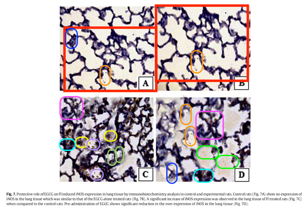


"Protective role of EGCG on Fl induced iNOS expression in lung tissue by immunohistochemistry analysis in control and experimental rats. Control rats (Fig. 7A) show no expression of iNOS in the lung tissue which was similar to that of the EGCG alone treated rats (Fig. 7B). A significant increase of iNOS expression was observed in the lung tissue of Fl treated rats (Fig. 7C) when compared to the control rats. Pre-administration of EGCG shows significant reduction in the over expression of iNOS in the lung tissue (Fig. 7D)."
The authors have taken steps to amend Fig 7, presumably directed through the journal, though they did not explain precisely how it found its way into [13]:
Dear professor, I have addressed the issue which u have mentioned the immunohistochemistry . I have submitted the new original picture after done the experint.
...I want to clarify one thing, in the figures that you have shown repeated elements were not created by us and it is naturally found in our histological slides. ... Repetitions that you have portrayed in the figures were not created by us and there was no need to manipulate in this particular panel of figures. ... Regarding immunohistological studies, the tissues were fixed and dispatched to the out sourcing lab for preparation of the slides using immunohistochemical staining and a pathologist would take the photograph from different groups and send their results with comments along the slides. ... I would give some of the published papers with these criteria, you can check it down with your so called figure plagiarism software. And i have reviewed more than 300 papers peer reviewed paper from around every publishing house and i have also evaluated the originality and correctness of every article to be published in the journals. By just going through the manuscript i could identify the originality of the articles even we are not having such sophisticated softwares and microscopes. ... And every journal system is having the iThenticate/Urkund softwares and other similar softwares for finding plagiarism and duplication/manipulation of materials/figures in the submitted manuscript. After these peer review process only our papers are accepted by the reputed journals. Do you want to disparage/disapprove all the systems, softwares and reviewers were inefficient and useless?

A charitable interpretation is that the images were originally labeled there, in a way that clashed with the journal's house style, obliging someone to paper over the labels with copy-paste because the original, unlabeled data files were no longer available.
There is a spectrum of manipulation. The intellects vast and cool and unsympathetic who occupy PubPeer could see no issue with most of the histology and immunohistochemistry images in the Miltonprabu oeuvre. Moving along the spectrum a little, we find alternations in the corners or more central, non-trivial but making no real difference to the message of each microphotograph. Below, Figs 5C and 5E from Thangapandiyan & Miltonprabu (2013) [2], and Fig 10 from [5].


Then there are overlaps, indicating that despite being labeled as coming from different conditions, two or three panels are sections of one single microphotograph. The tell-tale overlaps were small in Fig 9 from Thangapandiyan & Miltonprabu (2014) [4], and Fig 11 from Miltonprabu & Thangapandiyan (2015) [6].


Fig 11 from Thangapandiyan & Miltonprabu (2015) [9] made little effort to disguise its triple role.

Figs 5, 6 and 7 from Milton Prabu & Muthumani (2012) [1] are a wild triptych.

At the far end of the spectrum is this garish fabrication: an outcome of multiple cycles of sampling and re-mixing with overlaps as well, where any connection to real-world tissue samples is tenuous and fragmentary. Thangapandiyan et al. (2019) [18] were not well-served by the contractor in Figs 3 and 4. Perhaps the idea was to make the panels so painful to look at that reviewers wouldn't notice the malpractice.


To be fair to Miltonprabu and his colleagues, the journals' reviewers and editors did not detect these travesties either. And the problem should not arise in the future, because they have reduced their dependence on anonymous laboratories. To repeat the PP comment quoted above: "now we have equipped with our own photo microscopy, we didn't depend on the out sourcing fellows to take the photography our slides".
Any readers feeling unsated or inexhausted by my coverage are free to explore the relevant PubPeer threads themselves. In particular, I have skipped over the whole area of gel electrophoresis and Western blotting (Figs 6 and 7 from [16]).

And I was unsure whether these mirror-imaged and clone-stamped blood cells were optical or electron microscopy (Fig 9 from Nazima, Manoharan & Miltonprabu, 2016 [12]).

Sources:
[1]. "Silibinin ameliorates arsenic induced nephrotoxicity by abrogation of oxidative stress, inflammation and apoptosis in rats", S. Milton Prabu, M. Muthumani (2012).
Molecular Biology Reports doi: 10.1007/s11033-012-2029-6 [PubPeer].
[2]. "Epigallocatechin gallate effectively ameliorates fluoride-induced oxidative stress and DNA damage in the liver of rats", Shanmugam Thangapandiyan, Selvaraj Miltonprabu (2013).
Canadian Journal of Physiology & Pharmacology doi: 10.1139/cjpp-2012-0347 [PubPeer].
[3]. "Diallyl trisulfide (DATS) abrogates arsenic induced testicular oxidative stress in rats", Selvaraj Milton Prabu, Naorem Sumedha (2014).
International Journal of Pharmacology & Toxicology doi: 10.14419/ijpt.v2i2.2517 [PubPeer].
[4]. "Epigallocatechin gallate supplementation protects against renal injury induced by fluoride intoxication in rats: Role of Nrf2/HO-1 signaling", S. Thangapandiyan, S. Miltonprabu (2014).
Toxicology Reports doi: 10.1016/j.toxrep.2014.01.002 [PubPeer].
[5]. "Cadmium induced cardiac oxidative stress in rats and its attenuation by GSP through the activation of Nrf2 signaling pathway", Nazima Bashir, Vaihundam Manoharan, Selvaraj Miltonprabu (2015).
Chemico-Biological Interactions doi: 10.1016/j.cbi.2015.10.005 [PubPeer].
[6]. "Epigallocatechin gallate potentially attenuates Fluoride induced oxidative stress mediated cardiotoxicity and dyslipidemia in rats", S. Miltonprabu, S. Thangapandiyan (2015).
Journal of Trace Elements in Medicine and Biology doi: 10.1016/j.jtemb.2014.08.015 [PubPeer].
[7]. "Ameliorative efficacy of tetrahydrocurcumin against arsenic induced oxidative damage, dyslipidemia and hepatic mitochondrial toxicity in rats", M. Muthumani, S. Miltonprabu (2015).
Chemico-Biological Interactions doi: 10.1016/j.cbi.2015.04.006 [PubPeer].
[8]. "Cardiac mitochondrial oxidative stress and dysfunction induced by arsenic and its amelioration by diallyl trisulphide", Naorem Chanu Sumedha, Selvaraj Miltonprabu (2015).
Toxicology Research doi: 10.1039/c4tx00097h [PubPeer].
[9]. "Epigallocatechin gallate exacerbates fluoride-induced oxidative stress mediated testicular toxicity in rats through the activation of Nrf2 signaling pathway", S. Thangapandiyan, S. Miltonprabu (2015).
Asian Pacific Journal of Reproduction doi: 10.1016/j.apjr.2015.07.005 [PubPeer].
[10]. "Grape seed proanthocyanidins protects against cadmium induced oxidative pancreatitis in rats by attenuating oxidative stress, inflammation and apoptosis via Nrf-2/HO-1 signaling", Nazima Bashir, Vaihundam Manoharan, Selvaraj Miltonprabu (2016).
Journal of Nutritional Biochemistry doi: 10.1016/j.jnutbio.2016.03.001 [PubPeer].
[11]. "Hepatoprotective effect of grape seed proanthocyanidins on Cadmium-induced hepatic injury in rats: Possible involvement of mitochondrial dysfunction, inflammation and apoptosis", Selvaraj Miltonprabu, Nazimabashir, Vaihundam Manoharan (2016).
Toxicology Reports doi: 10.1016/j.toxrep.2015.11.010 [PubPeer].
[12]. "Oxidative stress induced by cadmium in the plasma, erythrocytes and lymphocytes of rats: Attenuation by grape seed proanthocyanidins", B Nazima, V Manoharan, S Miltonprabu (2016).
Human & Experimental Toxicology doi: 10.1177/0960327115591376 [PubPeer].
[13]. "Epigallocatechin gallate potentially abrogates fluoride induced lung oxidative stress, inflammation via Nrf2/Keap1 signaling pathway in rats: An in-vivo and in-silico study", Shanmugam Thangapandiyan, Selvaraj Miltonprabu, Poomalai Senthilraja (2016).
International Immunopharmacology doi: 10.1016/j.intimp.2016.07.022 [PubPeer]
[14]. "Diallyl trisulfide, a garlic polysulfide protects against As-induced renal oxidative nephrotoxicity, apoptosis and inflammation in rats by activating the Nrf2/ARE signaling pathway", S. Miltonprabu, N.C. Sumedha, P. Senthilraja (2017).
International Immunopharmacology (2017) doi: 10.1016/j.intimp.2017.06.011 [PubPeer].
[15]. "A mechanism underlying the neurotoxicity induced by sodium fluoride and its reversal by epigallocatechin gallate in the rat hippocampus: involvement of NrF2/Keap-1 signaling pathway", Thangapandiyan Shanmugam, Sharmilabanu Abdulla, Vadivazhagi Yakulasamy, Miltonprabu Selvaraj, Ramesh Mathan (2018).
Journal of Basic & Applied Zoology doi: 10.1186/s41936-018-0020-z [PubPeer].
[16]. "Sulforaphane potentially attenuates arsenic-induced nephrotoxicity via the PI3K/Akt/Nrf2 pathway in albino Wistar rats", Shanmugam Thangapandiyan, Mathan Ramesh, Selvaraj Miltonprabu, Tamilselvan Hema, Gunasekaran Bavithra Jothi, Venkatesan Nandhini (2019).
Environmental Science & Pollution Research doi: 10.1007/s11356-019-04502-w [PubPeer].
[17]. "The molecular and biochemical insight view of grape seed proanthocyanidins in ameliorating cadmium-induced testes-toxicity in rat model: implication of PI3K/Akt/Nrf-2 signaling", Nazima Bashir, Kalist Shagirtha, Vaikundam Manoharan, Selvaraj Miltonprabu (2019).
Bioscience Reports doi: 10.1042/bsr20180515 [PubPeer].
[18]. "Sulforaphane Potentially Ameliorates Arsenic Induced Hepatotoxicity in Albino Wistar Rats: Implication of PI3K/Akt/Nrf2 Signaling Pathway", Shanmugam Thangapandiyan, Mathan Ramesh, Tamilselvan Hema, Selvaraj Miltonprabu, Md Sahab Uddin, Venkatesan Nandhini, Gunasekaran Bavithra Jothi (2019).
Cellular Physiology & Biochemistry doi: 10.33594/000000082 [PubPeer].
No comments:
Post a Comment