
This post was earlier cross-posted at Leonid Schneider's site, hence the unfrivolous tone. The version there is improved by Leonid's editing and frame story.

I am in two minds whether Dong Ge Tong counts as another instance. Yes, his retraction notices explain that his implausible electron microscopy had been outsourced to a company that only retained the raw data for one month (while a laboratory flood destroyed the professor's own copies of the data files: plumbing skills are crucial for any serious scientist). But he had earlier credited the crystalline purity of the images in his papers to the diligence of an obsessive student.
#12 Dong Ge Tong
[...] If you have obsessive-compulsive disorder of symmetrical beauty, when you face such a vast and beautiful world, how would you like to express it? I bet you understand. The student has been doing the optimized experiments for two years, and as her teacher, I can only silently support her. If you have a student and she (or he) get a beautiful picture, have you asked your student if it was found, or is it the whole world? For high-resolution electron microscopy, you know, "finding" is the basic requirement. Luck is very important.
[...] It is obvious that we have either pasted the similar type of image for different material. This may have happened during outsourcing of our samples for characterization techniques. [...]
[...] Since We have no expertise in biology, We got in touch with biological institution. So, we are strongly apologizing for this unintentional error. Mostly, these type of errors happen due to the outsourcing images.[...]

"HRSEM images of (a) PEDOT, (b) Mg-HA,(c) PEDOT/Mg-HA coated 316L SS and (d) EDAX spectrum of PEDOT/Mg-HA bilayer coated 316L SS"
Let us continue with electron microscopy, for this is where Gopi and his colleagues were particularly ill-served by whoever provided them with imagery. Please compare at the left, Fig 3(c) from Gopi et al. (2013b) [3], and at the right, Fig 3(d) from Gopi et al. (2015e) [10].


They depict, respectively, porous Strontium-substituted or Sr/Mg-substituted HA coatings on surgical steel, with polypyrrone or poly(3,4-ethylenedioxythiophene as a kind of primer coat to promote adhesion. The textures are clearly different, while sharing visual motifs in common; one or other did not come directly from the electron microscope.


Figure 7 was reproduced in Gopi et al. (2016b) [22], as Fig 15.14. For Gopi's work on the chemistry and biology and physical characterisation of modified apatite has led to his recognition as an expert in the perennial genre of "green synthesis of [X] from botanical precursors". Editors invite him to reprise his discoveries in Review Chapters: in this case, under the title "Chemical and green routes for the synthesis of multifunctional pure and substituted nanohydroxyapatite for biomedical applications". This is convenient for our purposes as the chapter is a gateway into this section of his oeuvre.
Below, left: Fig 15.17, "SEM micrographs of the HA nanoparticles synthesized using sucrose as a chelating agent from natural and commercially available sources (a) commercial sucrose, (b) pineapple extract, (c) carrot extract, and (d) sugarcane extract." Who would have expected pineapple- and sugarcane-sourced sucrose to produce such similar nanoparticles, differing only in scale?


Above, right: Fig 15.19, "FESEM images of HA nanoparticles synthesized using different concentrations of pectin (wt.%): (a) 0, (b) 0.01, (c) 0.04, (d) 0.07, (e) 0.1, and (f) 0.15." Again, the scale changes but the nanoparticles are otherwise identical.
Finally, Fig 15.8 is reproduced from Fig 3 of Gopi et al (2014d) [9]. A sense of unreality pervades these scenes. Is it the ambiguous layering of picture planes, the uncertainty of illusory depth, reminiscent of Matta's cosmic dreamscapes? Here are its panels, annotated, not at all phopped:


But some of the tours de force in [9] did not find their way into that Review Paper [22]. Perhaps the authors felt the pressures of space. I feel less constrained in this post, so here are Figs 7(b) and 7(c), "Ca/Sr/Ce–HA nanoparticles synthesized with optimum concentration of Ce3+ (0.1 M) after soaking for ... (b) 14 and (c) 21 days in SBF solution":


Fig 4 consisted of "TEM images of (a) HA, (b) Ca/Sr–HA and (c) Ca/Sr/Ce–HA nanoparticles with optimum concentration (0.1 M) of Ce3+ synthesized by microwave irradiation method". Below left, 4(b). Below right, 4(a) is compared with Fig 8 from Gopi et al. (2014f), [11], where the particles are derived from banana-skin pectin.


The two images are almost the same, despite their different provenances and scale-bars. Yet they differ along the middle left edge, leading to the melancholy conclusion that one or other has been modified.

A second pie-slice provided Fig 12(e) in the same paper, "HRSEM cross sectional micrograph of 14 days immersion of P(EDOP-coEDOT)/M-HA-3 bilayer coated 316L SS in SBF solution" (below, left). What commends it to our attention here, apart from the repetitive nature of the polished substrate, is the discovery that the same flaky pastry topping also appeared atop a different substrate, in Fig 3(g) ("Cross-sectional micrograph of Sr-HAP/ZnO duplex-layer-coated AZ91 Mg alloy") of Gopi et al. (2014b) [6] (below at right).

An intermediate layer of 'M-HA-3" gravy also found its way, relabeled as a Polypyrrone-3 primer coat, into Fig 3(d) of [3]. The thickness of the crust looks to me like 3(d) is a shepherd's pie.

I could easily devote a post to Apatite pie-crust imagery, tracing the tradition back to Fig 11(b) in Gopi et al. (2013) [2], below at left. Perhaps it is an Artist's Impression of what "SEM cross-sectional micrographs of HAP coating on the post-passivated PoPD-coated 316L SS in Ringer’s solution.... b high magnification for the detection of passive film" would look like, if actually prepared.


Above right is Fig. 2 in Gopi et al. (2015) [13], "Cross-sectional SEM image of 0.5 wt.% H2O2 (optimum concentration) treated AZ91 magnesium alloy after 6 h of immersion in SBF solution".
An especially rewarding lode of Piecrust Pastiche is centred on Fig 3(d) of Gopi et al. (2015c) [16], "cross sectional SEM image of CNT/M-HAP coating on Ti".

The polished surface of the substrate, with its distinctively recurrent striations, had already appeared in Figures 1(e) of Gopi et al (2014b) [7], and 6(f) of Gopi et al (2014c) [8]. Note that the latter ("the cross sectional image of M-HAP coating on Ti-6Al-4V at 1 s pulse on time and 4 s pulse off time") has a double-layer crust, one layer being the piecrust from the former ("HAP coating on untreated ... Ti–6Al–4V alloy").

[Credit to Parvicirrus Dubius]
It appears a fourth time in Fig 3(e) of Chozhanathmisra et al. (2016) [20], which is beginning to resemble a Black Forest Gateau, though it has no interpretative legend.
This focus on the curiously striated substrate should not distract us from the topping of 3(d) of [16]. For we find it above a different pie filling - in a flourish of photoshop, with vertical duplications from internal rearrangement - as Fig 4(c) of Gopi et al. (2014) [5] ("cross-sectional image of ... the M-HAP coating on untreated Ti").


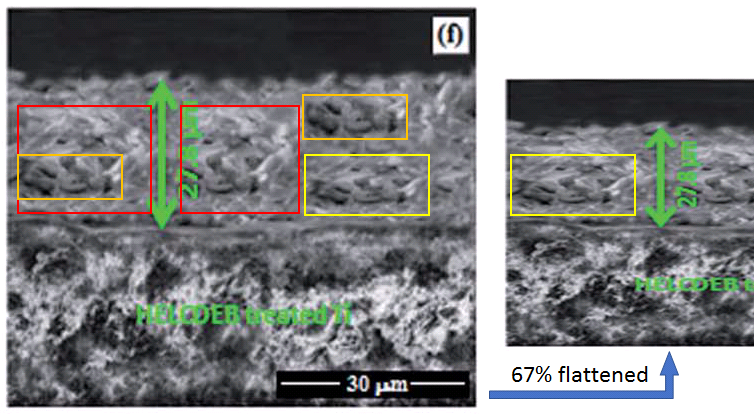
Still with [5], don't blink, or you will miss the transformation of 4(f) into 4(i) ("cross-sectional image of ... the M-HAP coating on 700 keV HELCDEB-treated Ti"). While 4(f) on its own ("cross-sectional image of ... the M-HAP coating on 500 keV HELCDEB-treated Ti") contains fractal graphic motifs deserving of your attention.

But at this point there is sad news to report... an Erratum has just been published for [24], replacing those iconic piecrusts in Figures 5(i) and 12(e) with less flamboyant photoshoppery. Evidently "they were inadvertently misrepresented" -- the outside consultants again. Rather than bicker and argue about who
Pastiche pies were not the only casualties of the Erratum, for there is more to biocompatible apatite coatings on metal implants than their cross-sections alone-. To assess their receptiveness to tissue regeneration, Gopi's team grew fibroblast cell-lines on the surfaces of the materials they synthesized. Then Figure 13 had to be replaced because its panels had already appeared as Fig 9 of [20], identified as "Optical images showing the viability of HOS MG63 cells on Zn-HNT/ M-HA bilayer coating for (a) control (b)1 (c) 4 and (d) 7 days" (rather than as "Optical images showing the viability of HOS MG63 cells on P(EDOP-co-EDOT)/MHA-3 bilayer coatings for (a) 1, (b) 4 and (c) 7 days of incubation").

They had also been identified as "HOS MG63 cells on S-PEEK/Sr-HAp composite coating for (a) 1, (b) 4 and (c) 7 days..." in Fig 8 of Rajeswari et al. (2014) [12] (below left); and as "HOS MG63 cells on control 4 (a,c,e) ... at 1 day, 4 days and 7 days of incubation" in Fig 6 of Gopi et al (2015b) [15] (below right).


I note in passing that regeneration is particularly good when fibroblast clusters are multiplied with the Photoshop clone stamp. Hence Fig 6 ("optical images of ... (c) CNTs/Sr-HAP,... (e) CNTs/Zn-HAP ... composite coatings on Ti obtained at 7 days of incubation") in Gopi et al (2015d) [17]... and then Fig 8.23 in Tiwari's book [21], for the authors were proud enough of these assisted results to reprint them.


Consolation can be taken from the fact that Fig 3 was not replaced, so presumably the authors have confidence in it. It is an X-Ray Diffraction pattern, artisanal and hand-made in nature, so we know that it was not created by graphical copy-paste. The diffraction peaks burst up from the baseline like Clavaria fungi, like zombies' fingers thrusting up through the cemetery soil as they dig their way out from their graves.

Fig 3 was the most recent in a series of diagrams in which the authors developed a new convention of what XRD patterns should look like, and whether a computer plotter or image file is really a requirement. Earlier zombie-finger exercises include Fig 3 from [12], and Fig 2 from [9].


Fig 2 of [6], and Fig 2 of [15]. Was the control profile meant to be a row of palm-trees?


Here by way of contrast are some more traditional diffractograms. At left, an early example: Fig 1 ("XRD patterns of HAP electrodeposited on 316L SS at (a) 1200 mV vs SCE, (b) 1400 mV vs SCE and (c) 1600 mV vs SCE") from Gopi et al. (2011) [1]. Duplicated stretches are marked. At right, Fig. 2 ("XRD patterns of (a) CNTs/HAP, (b) CNTs/Sr-HAP, (c) CNTs/Mg-HAP (d) CNTs/ZnHAP and (e) CNTs/M-HAP composite coatings") from [17].


The background of random zigzag noise is the same across the latter patterns, making it easy for one to hide behind another, for little distinguishes them except slight displacements along the Angle axis.


Those experimental outcomes must have been especially satisfying, for they appeared again (as original results) as Fig 8.19 of [21]; and earlier as Fig 2 ("XRD patterns for (a) HAP, (b) M-HAP and (c) CNT/M-HAP coatings") from [16] - shown below at left.


Above right is Fig 2 ("XRD patterns of (a) HA (b) M-HA (c) Zn-HNT and (c) Zn-HNT/ M-HA bilayer coatings") from [20], where the colors of (a) and (b) are all that distinguish them.

Fig 1 from Sathishkumar et al. (2016) [23] show "The FT-IR spectra of (a) HAP, (b) Sm/Gd-HAP-I, (c) Sm/Gd-HAP-II and (d) Sm/GdHAP-III coatings". I am not entirely comfortable with spectra that have overhangs. Were they left out in the sun?


The evidence suggests that the bactericidal performance of these coatings have felt the gentle touch of Appearance-Enhancing Software. The two Petri dishes in Fig 10 of [8] ("Photographs of antimicrobial test results of M-HAP coatings against (a) S. aureus and (b) E. coli (A - M-HAP coating obtained at 4 s pulse on time; and B - M-HAP coating obtained at 4 s pulse off time)") are more similar than one would expect. This is also true for Fig 6 from [12] ("Antibacterial activity of S-PEEK/Sr-HAp composite coating against (a) E. coli and (b) S. aureus, S-PEEK/Ce-HAp composite coating against (b) E. coli and (c) S. aureus and S-PEEK/Sr,Ce-HAp composite coating against (d) E. coli and (e) S. aureus bacteria").


Two last images. Fig 7 of Karthika et al. (2015) [19] showed "Petri dishes with MacConkey agar inoculated with (a) S. aureus and (b) E. coli, showing variable numbers of colonies when supplemented with different amounts of M-HAP/Gel nanocomposite". My own interpretation is that increasing the quantity of hydroxyapatite / gelatine combination improved the ability of agar to repel the Photoshop clone stamp, as seen more clearly with the magenta tint stripped away and the contrast enhanced.


AcknowledgmentsWith all the grants the Periyar team are receiving on the basis of this work, you would think they'd be able to afford more reliable outside consultants.
One of the authors D. Gopi acknowledges the major financial support from the Department of Science and Technology, New Delhi, India (DST) and Council of Scientific and Industrial Research, New Delhi, India (CSIR) in the form of major research projects. D. Gopi and L. Kavitha also acknowledge UGC, New Delhi, India for the Research Award (Ref. No. F.30-1/2013(SA-II)/RA-2012-14-NEW-SC-TAM-3240 and F. 30-1/2013(SA-II)/RA-2012-14-NEW-GE-TAM-3228) (2012-2014), respectively.
Sources:
[1]. "A facile electrodeposition of hydroxyapatite onto borate passivated surgical grade stainless steel", D. Gopi , V. Collins Arun Prakash , L. Kavitha , S. Kannan , P.R. Bhalaji , E. Shinyjoy , J.M.F. Ferreira (2011).Corrosion Science doi: 10.1016/j.corsci.2011.03.018 [PubPeer].
[2]. "Hydroxyapatite coating on selectively passivated and sensitively polymer-protected surgical grade stainless steel", D. Gopi , J. Indira , L. Kavitha , J. M. F. Ferreira (2013)
Journal of Applied Electrochemistry doi: 10.1007/s10800-012-0508-z [PubPeer].
[3]. "Corrosion protection performance of porous strontium hydroxyapatite coating on polypyrrole coated 316L stainless steel", D. Gopi, S. Ramya , D. Rajeswari , L. Kavitha (2013b).
Colloids and Surfaces B Biointerfaces doi: 10.1016/j.colsurfb.2013.01.065 [PubPeer].
[4] "A novel green template assisted synthesis of hydroxyapatite nanorods and their spectral characterization", D. Gopi, N. Bhuvaneshwari , J. Indira , K. Kanimozhi , L. Kavitha (2013c).
Spectrochimica acta. Part A, Molecular and biomolecular spectroscopy doi: 10.1016/j.saa.2013.01.052 [PubPeer].
[5]. "Investigation on corrosion protection and mechanical performance of minerals substituted hydroxyapatite coating on HELCDEB-treated titanium using pulsed electrodeposition method", D. Gopi , A. Karthika , D. Rajeswari , L. Kavitha , R. Pramod , Jishnu Dwivedi (2014).
RSC Advances doi: 10.1039/c4ra04484c [PubPeer].
[6]. "Electrodeposition of a porous strontium-substituted hydroxyapatite/zinc oxide duplex layer on AZ91 magnesium alloy for orthopedic applications", D. Gopi , N. Murugan , S. Ramya, L. Kavitha (2014b).
Journal of Materials Chemistry B doi: 10.1039/c4tb00960f [PubPeer].
[7]. "Evaluation of the mechanical and corrosion protection performance of electrodeposited hydroxyapatite on the high energy electron beam treated titanium alloy", D. Gopi, El-Sayed M. Sherif, D. Rajeswari , L. Kavitha , R. Pramod , Jishnu Dwivedi , S.R. Polak (2014b).
Journal of Alloys and Compounds doi: 10.1016/j.jallcom.2014.07.160 [PubPeer].
[8]. "In vitro biological performance of minerals substituted hydroxyapatite coating by pulsed electrodeposition method", Dhanaraj Gopi, Arumugam Karthika , Subramani Nithiya , Louis Kavitha (2014c).
Materials Chemistry & Physics. doi: 10.1016/j.matchemphys.2013.12.017 [PubPeer].
[9]. "Strontium, cerium co-substituted hydroxyapatite nanoparticles: Synthesis, characterization, antibacterial activity towards prokaryotic strains and in vitro studies", D. Gopi, S. Ramya , D. Rajeswari , P. Karthikeyan , L. Kavitha (2014d).
Colloids & Surfaces A: Physicochemical & Engineering Aspects doi: 10.1016/j.colsurfa.2014.03.035 [PubPeer].
[10]. "Development of strontium and magnesium substituted porous hydroxyapatite/poly(3,4-ethylenedioxythiophene) coating on surgical grade stainless steel and its bioactivity on osteoblast cells", D. Gopi, S. Ramya , D. Rajeswari , M. Surendiran , L. Kavitha (2014e).
Colloids & Surfaces B Biointerfaces doi: 10.1016/j.colsurfb.2013.10.011 [PubPeer].
[11]. "Novel banana peel pectin mediated green route for the synthesis of hydroxyapatite nanoparticles and their spectral characterization", D. Gopi, K. Kanimozhi , N. Bhuvaneshwari , J. Indira , L. Kavitha (2014f).
Spectrochimica acta. Part A, Molecular and biomolecular spectroscopy doi: 10.1016/j.saa.2013.09.034 [PubPeer].
[12]. "Investigation of anticorrosive, antibacterial and in vitro biological properties of a sulphonated poly(etheretherketone)/strontium, cerium co-substituted hydroxyapatite composite coating developed on surface treated surgical grade stainless steel for orthopedic applications", D. Rajeswari , D. Gopi, S. Ramya , L. Kavitha (2014).
RSC Advances doi: 10.1039/c4ra12207k [PubPeer].
[13]. "Evaluation of biodegradability of surface treated AZ91 magnesium alloy in SBF solution", D. Gopi , P.R. Bhalaji , S. Ramya , L. Kavitha (2015).
Journal of Industrial and Engineering Chemistry doi: 10.1016/j.jiec.2014.08.019 [PubPeer].
[14]. "Novel malic acid mediated green route for the synthesis of hydroxyapatite particles and their spectral characterization", D. Gopi, N. Bhuvaneshwari , L. Kavitha , S. Ramya (2015a).
Ceramics International doi: 10.1016/j.ceramint.2014.10.156 [PubPeer].
[15]. "Ball flower like manganese, strontium substituted hydroxyapatite/cerium oxide dual coatings on the AZ91 Mg alloy with improved bioactive and corrosion resistance properties for implant applications", D. Gopi , N. Murugan , S. Ramya , E. Shinyjoy , L. Kavitha (2015b).
RSC Advances doi: 10.1039/c5ra03432a [PubPeer].
[16]. "Single walled carbon nanotubes reinforced mineralized hydroxyapatite composite coatings on titanium for improved biocompatible implant applications", D. Gopi , E. Shinyjoy , A. Karthika , S. Nithiya , L. Kavitha , D. Rajeswari , Tingting Tang (2015c).
RSC Advances doi: 10.1039/c5ra04382d [PubPeer].
[17]. "Influence of ionic substitution in improving the biological property of carbon nanotubes reinforced hydroxyapatite composite coating on titanium for orthopedic applications", D. Gopi , E. Shinyjoy , L. Kavitha (2015d).
Ceramics International doi: 10.1016/j.ceramint.2014.12.114 [PubPeer].
[18]. "Fabrication of divalent ion substituted hydroxyapatite/gelatin nanocomposite coating on electron beam treated titanium: mechanical, anticorrosive, antibacterial and bioactive evaluations", A. Karthika , L. Kavitha , M. Surendiran , S. Kannan , D. Gopi (2015).
RSC Advances doi: 10.1039/c5ra05624a [PubPeer].
[19]. "Corrosion and biodegradability evaluation of magnesium substituted porous hydroxyapatite/polyethylene dioxythiophene bilayer coating on 316l stainless steel for orthopaedic applications", S. Ramya, D. Rajeswari, M. Palanisamy, D. Gopi , L. Kavitha (2015).
International Journal of ChemTech Research.
[20]. "Development of zinc-halloysite nanotube/minerals substituted hydroxyapatite bilayer coatings on titanium alloy for orthopedic applications", M. Chozhanathmisra , S. Ramya , L. Kavitha , D. Gopi (2016).
Colloids & Surfaces A: Physicochemical & Engineering Aspects doi: 10.1016/j.colsurfa.2016.10.018 [PubPeer].
[21]. "Carbon Nanotubes-reinforced Bioceramic Composite: An Advanced Coating Material for Orthopedic Applications", D. Gopi , D. Gopi , E. Shinyjoy , L. Kavitha , D. Rajeswari (2016).
Advanced Composite Materials doi: 10.1002/9781119242666.ch8 [PubPeer].
[22]. "Chemical and green routes for the synthesis of multifunctional pure and substituted nanohydroxyapatite for biomedical applications", Dhanaraj Gopi , Louis Kavitha , Subramanian Ramya , Durairajan Rajeswari (2016b).
Engineering of Nanobiomaterials doi: 10.1016/b978-0-323-41532-3.00015-4 [PubPeer].
[23]. "Tailoring the Sm/Gd-Substituted Hydroxyapatite Coating on Biomedical AISI 316L SS: Exploration of Corrosion Resistance, Protein Profiling, Osteocompatibility, and Osteogenic Differentiation for Orthopedic Implant Applications", Saravanan Sathishkumar , Kavitha Louis , Elangomannan Shinyjoy , Dhanaraj Gopi (2016).
Industrial & Engineering Chemistry Research doi: 10.1021/acs.iecr.5b04329 [PubPeer].
[24]. "Fabrication of Minerals Substituted Porous Hydroxyapaptite/Poly(3,4-ethylenedioxy pyrrole-co-3,4-ethylenedioxythiophene) Bilayer Coatings on Surgical Grade Stainless Steel and Its Antibacterial and Biological Activities for Orthopedic Applications", Ramya Subramani , Shinyjoy Elangomannan , Kavitha Louis , Soundarapandian Kannan, Dhanaraj Gopi (2016).
ACS Applied Materials & Interfaces doi: 10.1021/acsami.6b01795 [PubPeer].
No comments:
Post a Comment