
This post was earlier cross-posted at Leonid Schneider's site, hence the nonfrivolity. The version there is improved by Leonid's editing and explanation of the back-story.

The following post dwells yet again upon curious phenomena encountered in nanotechnology journals. This is becoming a regular theme. Readers may be hoping for something especially outrageous about the present investigation, but life is full of disappointments. At least there are these charming images of luminescent deep-sea fish in the abyssal dark-field.

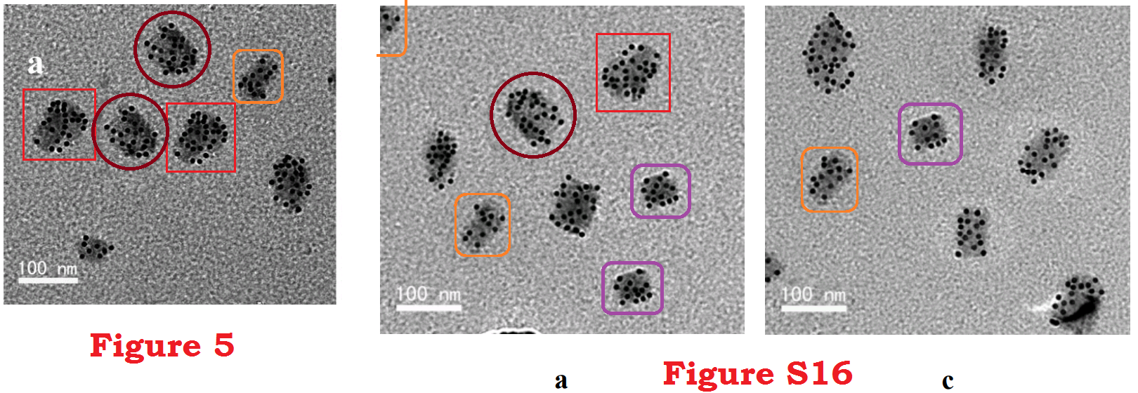
Alternatively, they are fragments of carbon (in some unspecified phase) bedizened with nanocrystals of Rh2Ni. The way that each nanocrystalline arrangement recurs across multiple carbon flecks, and indeed across more than one habit of nanocrystal, can be ascribed to Sheldrake's Morphogenetic Field. As can the flecks' resemblance to Terry Gilliam drawings.
In "A Different Universe" (2008), physicist Robert P. Laughlin had some thoughts that shed a light on the state of the nanotechnology literature (or "advanced-materials" if you like):
"About halfway through the daylong program of my first review I saw a presentation by our electron microscopist that knocked my socks off. [...] Her presentation was less a technical seminar than a National Geographic special on the Escalante staircase or the Himalayan foothills of Tibet. She showed a sequence of the most astonishing topographies, no two of which were the same. [...] Then came a forest of grotesque gargoyles on stems planted at the edges of lakelike dimples, thirsty cauliflower creatures from another planet descending on New England to seek out ponds. After that a formidable mountain range with strange caps on the top resembling snow, as one might see from an airplane flying over Aspen or Katmandu."Within this literature of interest, we find a series of papers from a research group in Chengdu, Sichuan. Interest in them has blown up rapidly over just a few days, with no end of Tweeted incredulity, and a Reddit discussion, as well as the inevitable PubPeer threads that supplied most of the raw material for this post. So perhaps there is still some lingering relevance in the topic of nanojunk, and I am not simply flogging a dead low-hanging fruit in a barrel.
"At the scale visible to the electron microscope, every surface looked interesting. [...] great talent would be required to take a dull electron micrograph of a surface."
"The structures displayed by my colleague the electron microscopist are prototypical of what I call nanobaubles, fascinating and beautiful structures that develop spontaneously at small scales but have no known use except as entertainment."
The earlier papers in this series appeared in journals from Elsevier and Springer, with unexceptional Impact Factors like 2.993 (Journal of Materials Science) and 2.687 (Materials Science). Following the "nano-compost" tradition, they announced such developments as antibacterial CuO2 nanosponges derived derived using pine-needle templates, and photocatalytic "biomorphic nickel oxide microtubes" derived from cotton through calcination, molecular substitution, and the full panoply of alchemical transformation. Promising superior battery performance, and hydrogen generation from catalysed photolysis of water, and superior pollutant detection, these papers were typical of the genre and in line with Professor Laughlin's summary.
"...we dream to engineer new kinds of useful gadgets and products for practical use, such as early warning sensors for obnoxious smells or machines that render leftover banana peels into gasoline."But at some point the authors grew in ambition (or drew down a large research-project grant), for they switched to targeting Royal Society of Chemistry journals like J. Mat. Chem. A and Nanoscale (and ACS Applied Materials & Interfaces from the American equivalent), with three times the Impact Factor: IF = 9.931 and 7.233 respectively (and 8.097). One expects the peer-reviewing there to be correspondingly more stringent and demanding. Anyway, that is what raised these papers to the threshold of attention. For these august journals have brought us implausible images like these:

Then it emerged that four of these papers recently and concurrently attracted Expressions of Concern from the editors of J. Mat. Chem. A and Nanoscale, "in order to alert our readers to the fact that we are presently unable to confirm the accuracy of the data reported in the TEM images". The concern focussed on the STEM imagery, which calls out for verification. Apparently the original data files are unavailable in each case, so we read (repeatedly) that
The authors are in the process of repeating the experiments to confirm the validity of the TEM images in the published Figures. This notice will be updated when a conclusive outcome is reached.These four papers:
1. Monodisperse Ni3Fe single-crystalline nanospheres as a highly efficient catalyst for the complete conversion of hydrous hydrazine to hydrogen at room temperature;
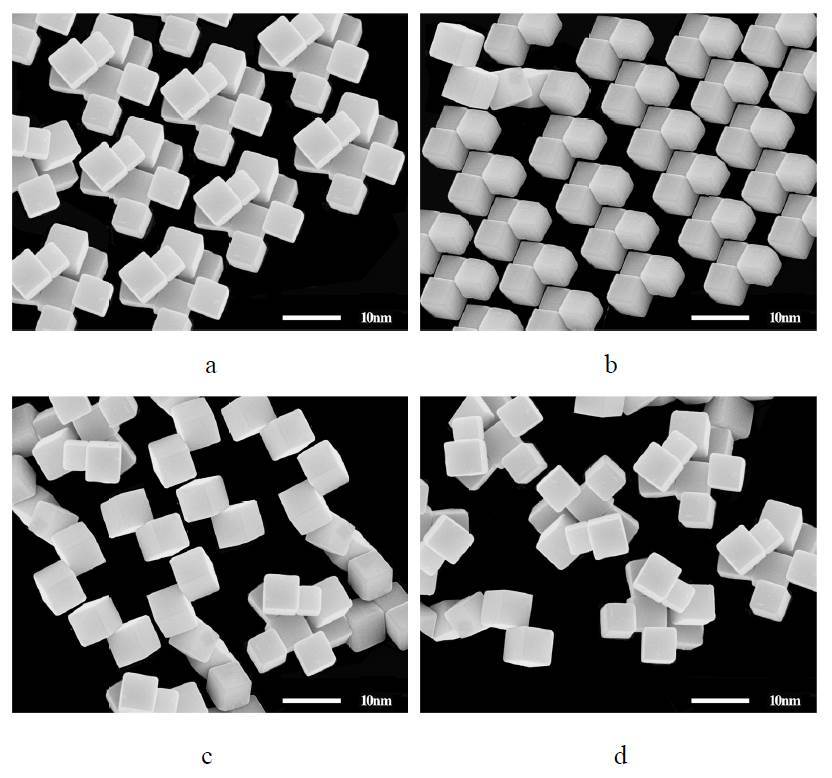
2. Preparation of face-centered-cubic indium nanocubes and their superior dehydrogenation activity towards aqueous hydrazine with the assistance of light;

3. Synthesis of octahedral, truncated octahedral, and cubic Rh2Ni nanocrystals and their structure–activity relationship for the decomposition of hydrazine in aqueous solution to hydrogen (the source of the luminescent fish);

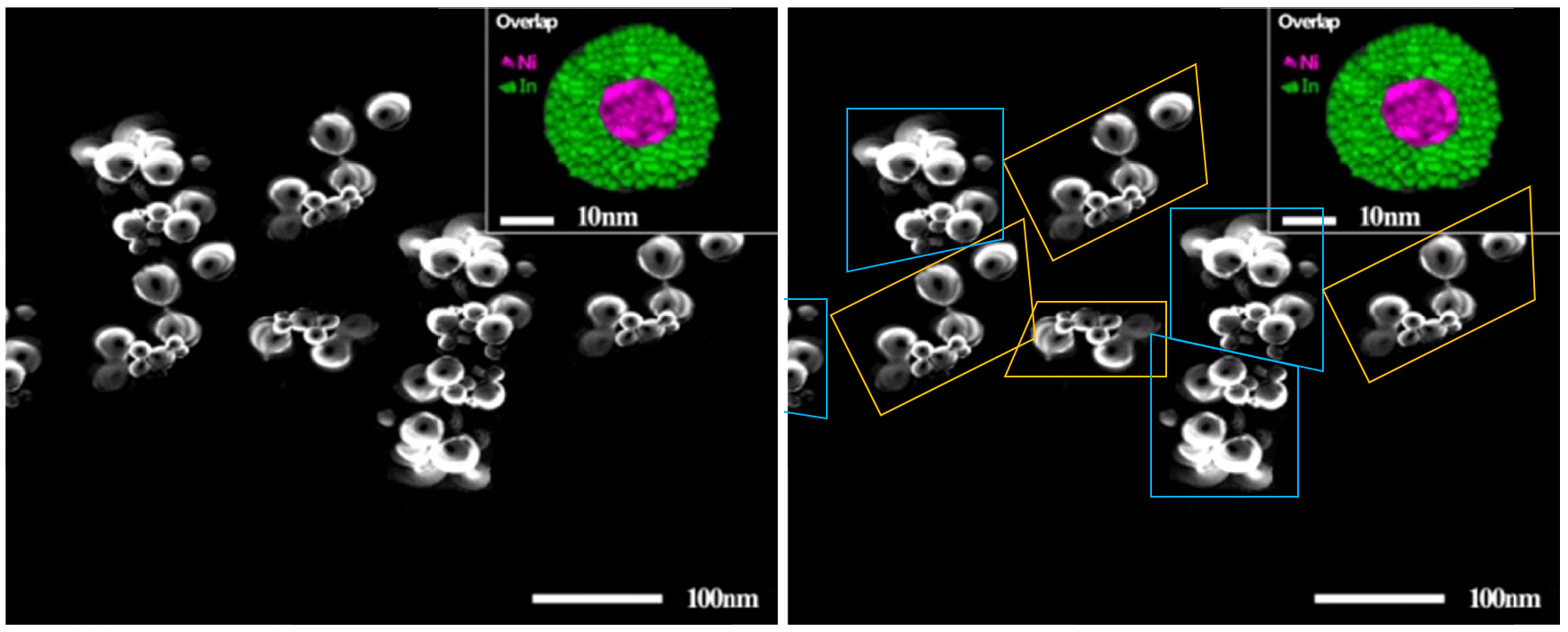
4. Hollow amorphous NaFePO4 nanospheres as a high-capacity and high-rate cathode for sodium-ion batteries.

Dong Ge Tong (the group's leader) responded in a couple of PubPeer threads to chide other commenters for their skepticism, and their inexperience with experimental observation.
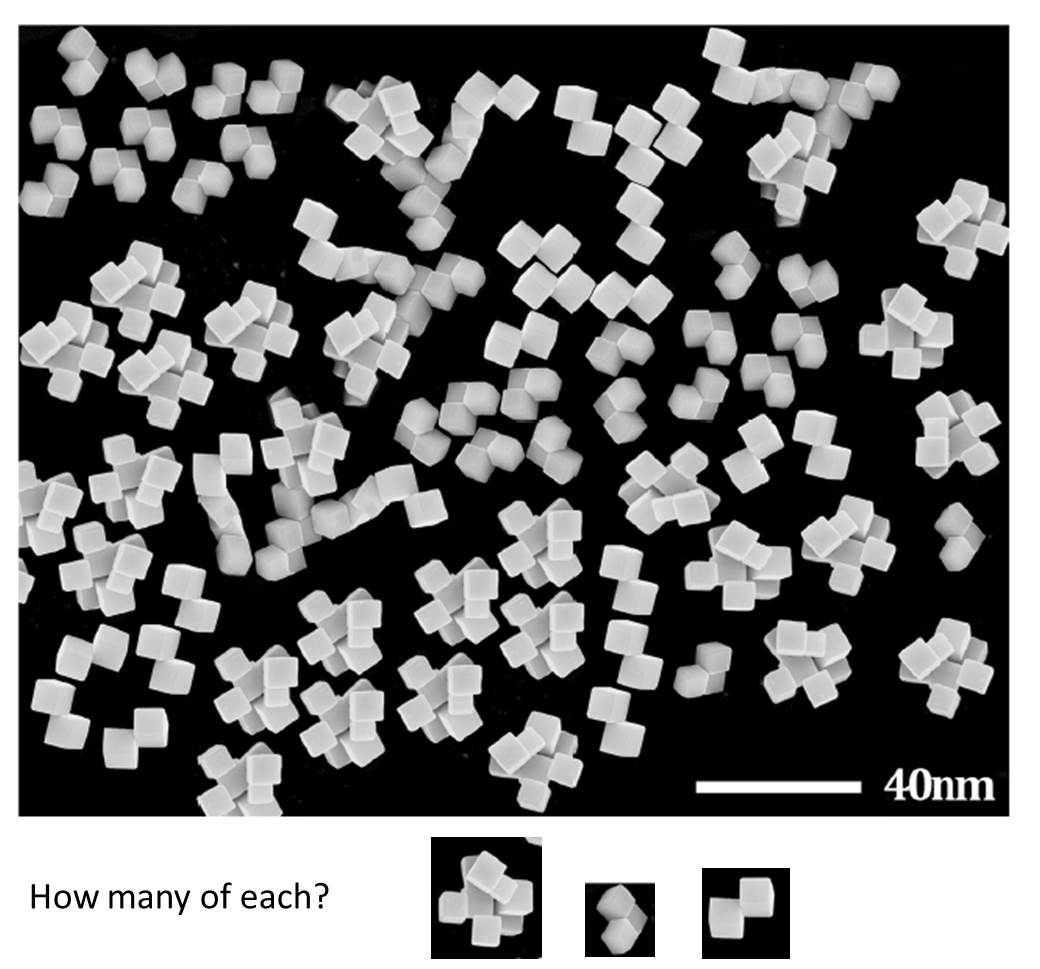
The repetitive elements in the figure for a optimized sample under an electron microscope is normal. Thus, I sincerely hope you do an experiment yourself with a good sample to observe the repetitive elements under an electron microscope, rather than using computer graphics software to evaluate it. For a good sample, it can be see that there are lots of repetitive elements. Moreover, with some special dispersing addition, some special order you can see.It appears that images of geometrical clarity are within the grasp of anyone who has the patience to scan enough of the infinite universe of the Nanocosm and select the most aesthetic results.
The world observed by the electron microscope is very large, but we can only record or present a small part of them. Whether this part of the record can scientifically reflect the real world observed by electron microscopy has become a matter of debate. Some people think that they should be unsuitable at the same time. Just like you, I agree with you very much. However, in real experiments, because of the problems of sample preparation and equipment itself, it is possible to see the disagreeable places and the agreeable places together under the lens, but can not take them all together. And when you use one of them, you will select the one which is very pleasing to your eye. It is just like that when you're looking for a job, you will use your favorite ID. But, you are the same as you. At the same time, I know you are a good person. Thank you very much for your comment.So there is a corner of the Nanocosm full of googly eyes:
It contributes to thread #5, "Mesoporous Face-Centered-Cubic In4Ni Alloy Nanorices: Superior Catalysts for Hydrazine Dehydrogenation in Aqueous Solution" (so far without Editorial comment). I was disappointed, by the way, to discover that "nanorices" is not the plural of "nanorix", but simply a fresh coinage for Phop-cloned nanoparticles inspired by short-grain rice.
Thread #7 is Monodisperse CuB23 nanoparticles grown on graphene as highly efficient catalysts for unactivated alkyl halide Heck coupling and levulinic acid hydrogenation. This corner of the Nanocosm is devoted to exercises in decoupage - cutting out different shapes from the same sheet of nanoparticle-adorned graphene. With one outline, the sheet becomes a dog's head, while another turns it into a left-facing profile of Elvis (as played by Bruce Campbell).

 The wording of those four Concerned Expressions (and any more that are still to come) leaves two possibilities open. The authors might have been invited to repeat the observations, and to replace the contested data-free Figures with better-documented substitutes (and if serendipitous Soma-cube configurations of nanocubes are really so frequent, this should not be difficult)... or they might have offered to do so, with the editors accepting the offer in the interests of a fair process. But the sudden appearance of four Notices, unprovoked by any obvious harassment from outsiders, suggest that the RSC publishing arm has belatedly lost tolerance for CGI and Photoshop as substitutes for actual microscopy.
Anyway, we learn from Dr Tong that the credit for exploring the Nanocosm through electron microphotography belongs to a well-trained and obsessive-compulsive student:
There is a disease in the world called obsessive compulsive disorder, like Holmes in the movie. The world is so large under the electron microscope that if you want, you can find two identical ones. Electron microscopy is also called aesthetics, and how you would like to express the world you see. If you've done the experiment, you know there's a word in the electron microscope experiment called "find", to find what you want to say. So, sincerely hope you to do a good sample and bad sample of the electron microscope experiment comparison, you will understand what is lucky, what is incredible. If you have obsessive-compulsive disorder of symmetrical beauty, when you face such a vast and beautiful world, how would you like to express it? I bet you understand. The student has been doing the optimized experiments for two years, and as her teacher, I can only silently support her. If you have a student and she (or he) get a beautiful picture, have you asked your student if it was found, or is it the whole world? For high-resolution electron microscopy, you know, "finding" is the basic requirement. Luck is very important.Student, meet bus.
Problematic aspects are not confined to the STEM Figures. A recurring feature of these papers are X-Ray Diffraction profiles in which a 'flatline' section of the pattern turns out on close inspection to consist of a single shorter rusty-sawblade segment, repeated several times over. In #6:
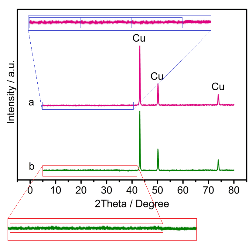
The coincidences continue to accumulate when one of these synthetic-flatline XRD profiles is repeated (down to the level of pixels of noise) and presented as the result of several independent but well-replicated experiments. From #1:
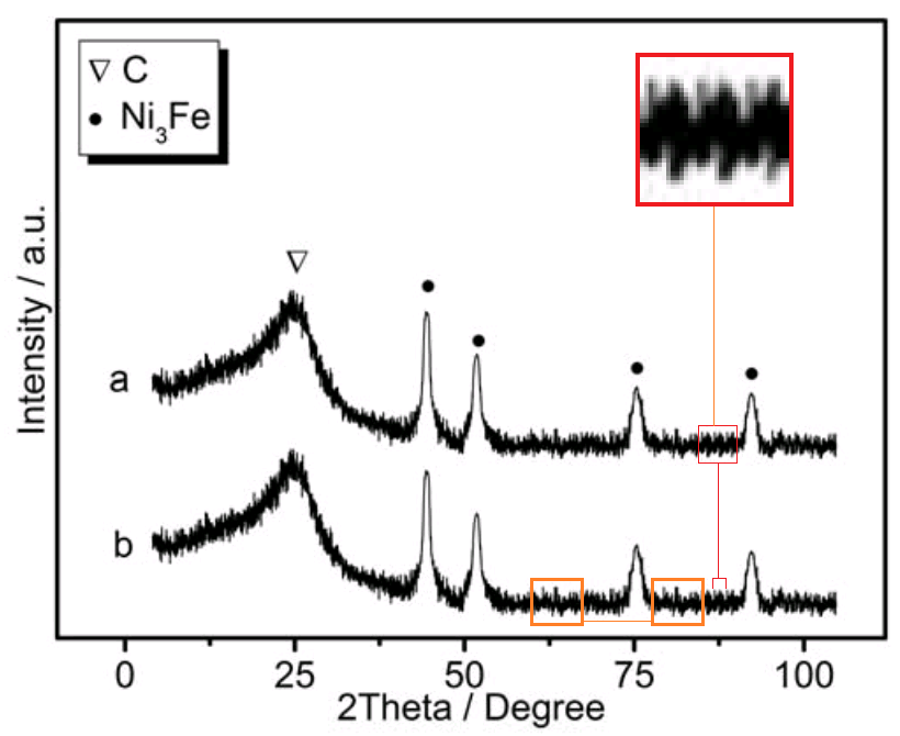
From #7:


I leave the last word to Dr Tong.
We respect you very much. You are all good people. Because of our limited knowledge, in fact, some experimental results we do not understand enough until now, just only to think and apply them simply. These results may include other useful information. Yours valuable comments will help us to think and understand the experimental results more deeply. Thank you very much for your comments. Thanks a lot.
------------------------------------------
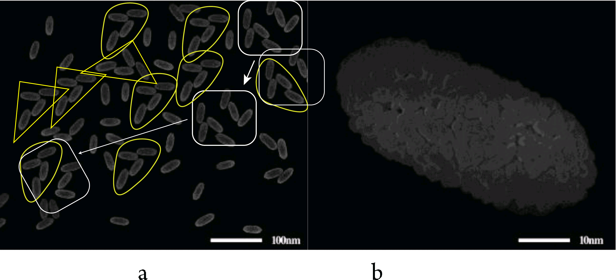
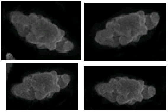
Afterthoughtage #1: Dead-horse-flogging activities have continued in the Pubpeer threads. In the Nanorice-grain paper, many of the Supplementary Figures purport to show the development of Nanorices from different chemical mixtures… in fact they are collages worthy of Hannah Höch, based on random SEMs of textured surfaces that have been cut up, duplicated and rearranged.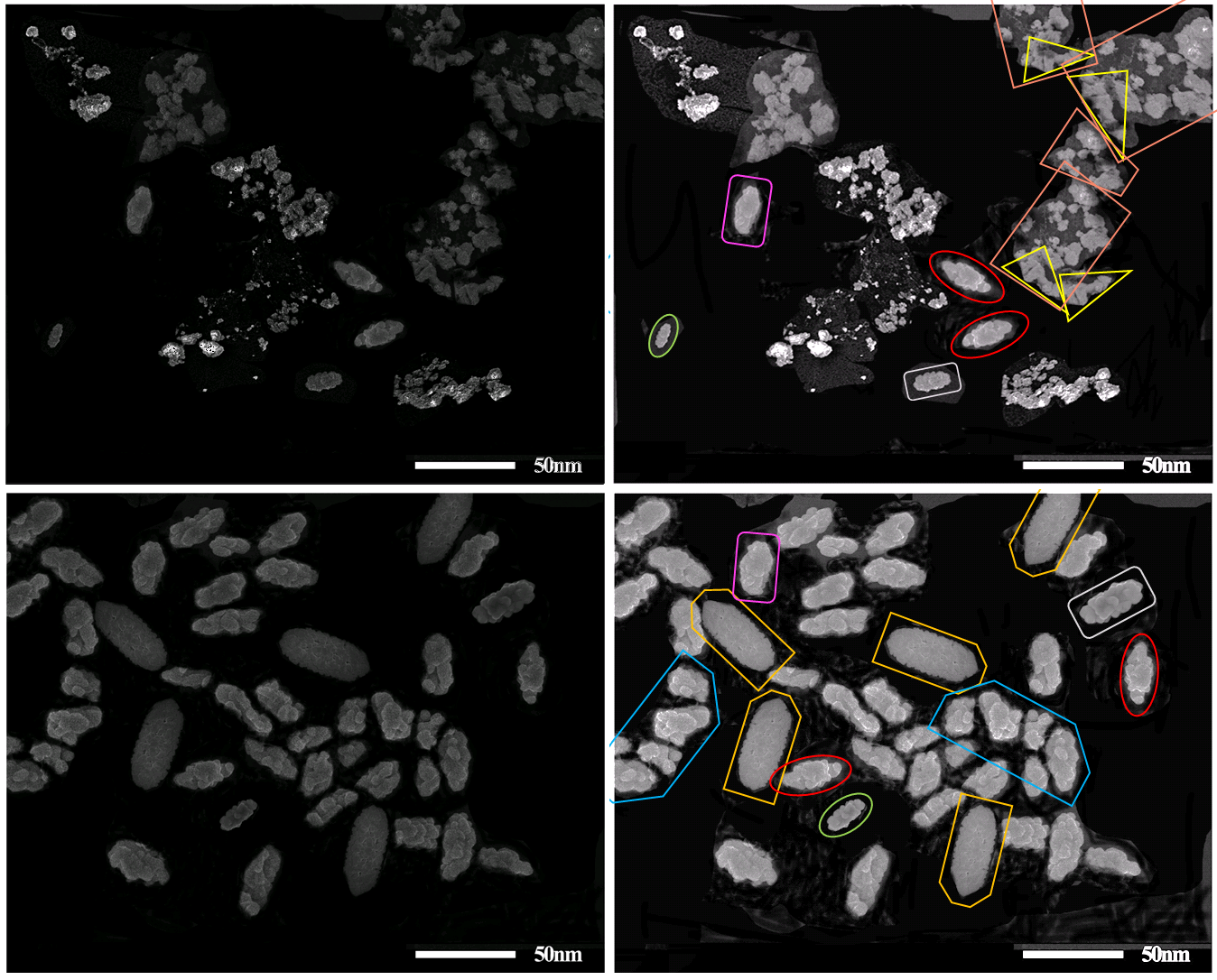
Some of the repeating pictorial elements in these collages have been vignetted and turned into oval ‘particles’ by darkening the background around them:

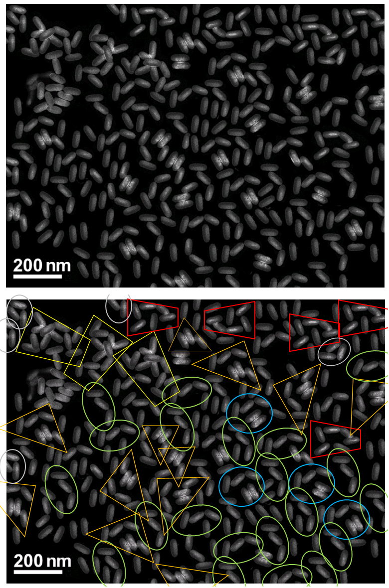
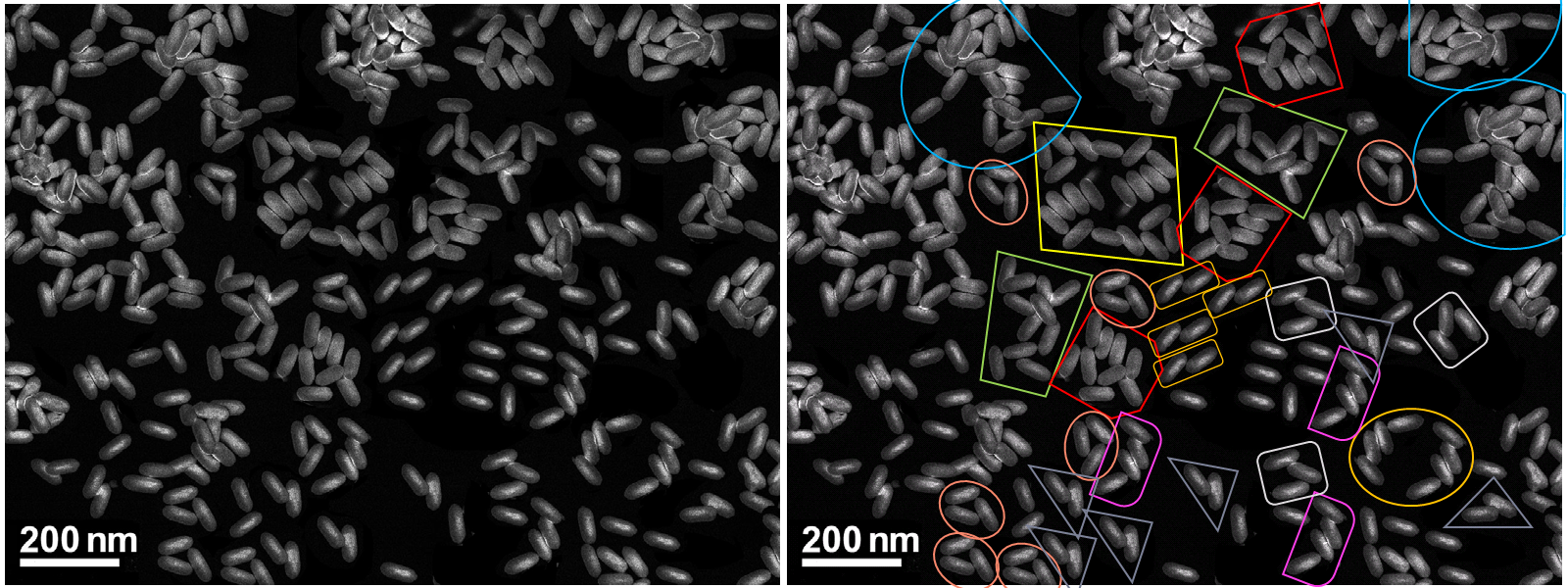
Afterthinking #2: The ‘nanococoon’ Supplementary Figures are also full of examples of SEMs of textured surfaces, divided up into nascent nanoparticles by drawing dark lines between them with a Photoshop crayon.
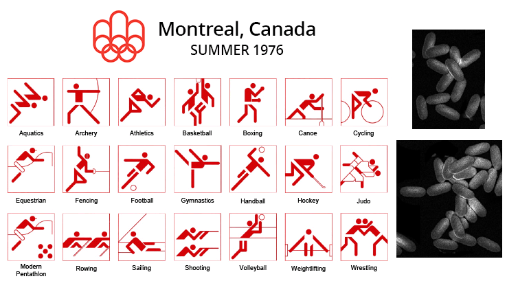
Were some of the arrangements of imaginary particles intended as a homage to Olympic sports pictograms? It would be irresponsible not to speculate.
No comments:
Post a Comment