
This post was earlier cross-posted at Leonid Schneider's site, hence the nonfrivolity and Explaining Voice. The version there is improved by Leonid's editing and explanation of the back-story.

Just when you were getting used to 'liposomes' and 'exosomes' entering the advertising lexicon of cosmetics and health quackery as signifiers of Sciencyness, along came niosomes and transfersomes and vesosomes and emulsomes. Anyone who identified these as "The cows from Cold Comfort Farm" has failed the test (the judges' decision is final).
In fact these are all additions to the menagerie of Drug Delivery research: different ways of sequestering a drug within a microscopic or nanoscopic vesicle with the idea of delivering the molecules to a specific tissue instead of wasting them all over the body. The drug might be encapsulated within cubosomes, for instance, in a form that can be snorted, then transported to the brain along olfactory neurons, thus dodging the blood-brain barrier. Because Drug Delivery straddles the boundary between nanotechnology and molecular biology, papers can feature faked diagrams from both fields - photoshopped electron microscopy and immunohistochemistry. So far "noisome", "handsome", "toothsome", "quarrelsome" and "meddlesome" are not recognised as Drug Delivery terms, and now my ambition is to publish a paper introducing them all.
With that as context, here to pique the readers' interest are Figures 7 and 6 respectively from Jain et al (2012) [A2] and Choudhary et al (2016) [B12].


They are filigree pastel crystals, yet somehow woven or crocheted as well. One can only be sure that there is nothing biological about them, even though the figure captions identify them as the stomach mucosa of rats, bearing alcohol-induced peptic ulcers: treated in [A2] with ranitidine bismuth citrate and amoxicillin trihydrate (delivered by concentric nested liposomes) and in [B12] with rabeprazole and amoxicillin (delivered by "polymeric low-density microballoons"). The two papers have Govind P. Agrawal as a shared author.


What is one to make of Figures 1 of Rai et al (2016) [B11] and 1 of Sudheesh et al (2011) [C17]? The former depicts "Eudragit-coated dextran microspheres of 5-fluorouracil" (targeting the colon) while the latter are "tetanus toxoid-loaded aminated gelatin nanoparticles" (for vaccines). Close inspection reveals the 2011 version to be a swarm of replicated pairs and triplets of spheres. Yet these Bubbles Congeries [Lovecraft 1931] have enough in common besides the background that their single origin is undeniable.

As the head of the latter laboratory, a Dr S. P. Vyas responded on the PubPeer thread for [C17]. Alas, his answers revealed little, except that he is not accustomed to questions... not even to questions of a rhetorical nature, that are asked into the void and not directly at him. Also, it is below his dignity to haggle with anonymous critics, although onymous critics are equally beneath his contempt.
#3 S. P. Vyas
Why should we explain this to you?
#6 S. P. Vyas
We wrote the paper. If there is a mistake, we can choose to correct it or not. We don't think pubpeer is the right place to respond to anoymous people.
No-one can accuse the ADINA Institute of wasting research funds. Once they have created an image it receives maximum mileage. In a trilogy of papers, a Scanning Electron Microscopy (SEM) Phop variously depicts microballoons with payloads of Famotidine for the upper small intestine (Fig 3B, Choudan et al 2013 [A3]), or of Mesalamine chloride (1B, Jain & Jain 2013a [A4]); or perhaps they are phenylalanine-anchored liquid nanocarriers to treat AIDS-related encephalopathy (3B, Vyas et al 2015 [A9]). SEMs like this will be a recurring theme through the rest of this post. TEMs will be another.


These multiwalled carbon nanotubes conjugated with hyaluronic acid (to target colon cancer) were only used once, in Prajapati et al (2019) [A10]. But one cannot question the team's Photoshop skills. I recognise 3(a) as a homage to the back-cover album art for "Dance of the Lemmings".


Figures 7 from Bilthariya et al (2015) [A8] and 8 from Jain et al (2014) [A6] show tissue fluorescing after permeation with FITC. The only uncertainty is whether the tissue came from "inflamed joint sac" or "liver sac", and whether its entry into the cells was facilitated by particles of folate-conjugated albumin or lactosaminated-N-succinyl chitosan, laden with etoricoxib or acyclovir.

[A6] and [A7] are also a convenient gateway to Figs 5(b) and 4(b), where the reason for manipulating one or other image (or both!) is unclear; perhaps it was for practice, or to create a Spot-the-Difference puzzle


Fluorescent tissue samples is becoming another recurring theme in this post. Below, Figs 4 from Jain, Agrawal & Agrawal (2011) [A1], and 3B from Jain & Jain (2013b) [A5]. Both are a "fluorescence photomicrograph of rhodamine-123 labeled nanolipobeads formulation", designed to cure stomach ulcers by eliminating H. Pylori. The 2013 version was photoshopped with the triplicated nanolipobeads at the right either to defuse accusations of self-plagiarism, or because the authors really like the design aesthetic of 1970s Krautrock album art.


It is almost time to move on. To include everything covered in the relevant PubPeer threads would be exhaustive even by my standards, so out of great-souled mettā I have organised the haul from trawling those threads into a spreadsheet for interested readers to consult. The spreadsheet (like the Sources section at the bottom of this post) is divided for convenience into three sections, but boundaries are permeable:A.K. Jain was still affiliated to Dr H.S.Gour University for the first five of the 13 papers grouped together under his name, not yet having assumed leadership of the ADINA institute, and G.P. Agrawal (Jain's colleague or mentor, I assume) was co-author on those five. The two institutions enjoy a close, image-sharing association. There is no better way to illustrate this collegiality than Fig 5C of [A5], which had earlier appeared as Fig 4 of Jain et al (2009a) [C7] - filed below in the "Suresh P. Vyas" section.

[H/t Sebastes Hopkinsi!]
The pedants and blackboard monitors and incorrigible scolds who hang out at PubPeer have devoted 15 threads to Dr Agrawal's papers, excluding the five co-authored with Jain, but including five co-authored with S. P. Vyas. My generosity is boundless and I created a diagram to help keep track of the co-authorship situation.

We have already seen one repurposed / modified image, the "Bubbles Congeries" shared between [B11] and [C17]. To continue in this vein, here's a SDS-PAGE blot (a form of protein-separating electrophoresis) showing how a nasal vaccine against Hep-B works by wrapping up HBsAg surface antigens within liposomes. More specifically, within "Immunoglobulin immobilized liposomal constructs" in one incarnation (Fig 4 from B. Tiwari et al 2011 [B7]) and within "Viral protein complexed liposomes" in the other (Fig 3 from S. Tiwari et al 2011 [B8]).

These fluorescing cell clusters demonstrate how HNGC1 tumor-cell clusters absorb Methotrexate better (or perhaps doxorubicin) when the drug is concealed inside Cationized Albumin Conjugated Solid Lipid Nanoparticles (or perhaps in Cationic ligand appended nanoconstructs), thus curing brain cancer. In Fig 13 of Agrawal et al (2011a) [B4], the cells are multiplied by zooming in on them at three different sizes.

In Fig 11 of Agrawal et al (2011b) [B3], the same cells pair up with 180°-flipped copies of themselves.

In general the Pharmaceuticals Research Laboratory staff are profligate with digital resources and their photoshops are not recycled so thoroughly. The next examples are one-offs. Boxes mark the cloned sections in Fig 7(a) of [B3], a TEM photomicrograph of SLN-DOX.

Much could be said about the thumb-print Solid-Lipid Nanoparticles (plain and CBSA conjugated) in Fig 5 of [B4]. It is easier, though, to bring up the wallpapered backgrounds with contrast enhancement:


In Fig 1(b) of [B7], the cluster at lower right reminds me of a stylised figure fleeing the scene after duplicating the liposomal constructs at centre. Enhanced contrast calls attention to glitched repeating margins around the lower-left corner.

Figs 2 of Yadav et al (2010) [B2] and 2 of Jain et al (2014) [B10] fell victim to extreme image compression at some stage, turning them into seas of JPG artefacts. The lack of resolution makes it difficult to comment on any similarities among the nanoparticles ("hyaluronic acid decorated PLGA nanoparticles for delivery of 5-fluorouracil" and "Adapalene loaded solid lipid nanoparticles gel" for acne treatment respectively), but areas of background have been copy-pasted.


Please be patient for one more example. In Jain et al (2010) [B1] the Solid Lipid Nanoparticles were mannosylated for better delivery of doxorubicin to brain tumours. The backgrounds of Figs 1(a) and (b) are surprisingly similar, while a 'T' motif -- a kind of watermark, perhaps -- repeats across 1(b).

The outlines of the NPs themselves only repeat in a few pairs and triplets. Still, it is clear from similarities among their internal structures that they were cut out from a handful of templates, as exercises in decoupage worthy of Matisse.

The PubPeer thread for [B1] came to the attention of the authors, with the lead / corresponding author replying first, before responsibility shifted to a minor co-author.
#2 Himanshu Agrawal
Yes there is a mistake! Thank you for pointing out.
#5 Saikat Majumder
We thank Dr Hoya and Dr Actinopolyspora for pointing out these serious errors in our published articles. we will carefully examine the whole data. If we find the data are incorrect,we will redo the experiment and then publish the correct data. If we are unable to repeat the experiments,we will contact the journal immediately and ask for retraction. Thank you again for reminding us these concerns!

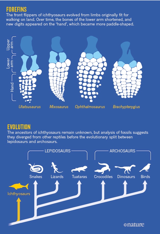
Fig 5 of [B5] featured these views of the NPs adhering to Peyer's Patches in mouse intestine, though a comparison of the green nebulae of fluorescing NPs reveals A and B to be tailored versions of a single original. They are not false-color x-rays of ichthyosaur flipper bones.

Thus the reappearance of these frames in Fig 7 of [C23], now as chitosan NPs, are four uses of a single image. The Indian Council of Medical Research must be pleased by such thrift in the use of their research grants.


At this point the comparison between Figures 1 of Paliwal et al (2011) [B6] and 1D of Paliwal et al (2013) [C8] is invidious, but I cannot resist the temptation to flog a dead horse with another dead horse. In [B6] the Solid-Lipid NPs are biomimetic and laden with a payload of lipid-conjugate low-molecular-weight heparin; in [C8] they are glyceryl behenate-cored and laden with methotrexate; perhaps that is why there are fewer of them.


"Dual targeted polymeric nanoparticles based on tumor endothelium and tumor cells for enhanced antitumor drug delivery" (Gupta et al 2014 [B9])? Or "lipidic nanoparticles for dermal delivery of fluconazole against cutaneous candidiasis" (Gutpa & Vyas 2012 [C19])?


Anyway, on to Dr Sureth P. Vyas. As one mark of his productivity, his H-Index is 64, and those 50 papers addressed in PubPeer threads span only some 10 years of his career.
Three of those papers were from a veritable scientific supergroup: the coauthors included Kailish C. Gupta, erstwhile Director of the CSIR-Indian Institute of Toxicology Research and no stranger to PubPeer in his own right. The last I checked, an inquiry into the research methods of Dr Gupta and others at CSIR-IITR had finished and a report was awaiting action... that was in January but people can be forgiven if events have distracted them. My point is that we have reached the highest levels in the Indian research establishment.


Pathak et al (2009a) [C10]:

Now [C10] and Pathak et al (2009b) [C9] addressed, respectively, the tumor targeting of "polysaccharide decked polyethylenimine based nanocomposites" and tumor gene therapy mediated by "Chondroitin Sulfate−PEI Nanoconstructs", with surprisingly similar results.


https://www.dovepress.com/journal-editor-integrated-pharmacy-research-and-practice-eic135
https://www.journals.elsevier.com/international-journal-of-pharmaceutics/editorial-board
https://www.future-science.com/journals/tde/editors
https://scienceforecastoa.com/Journals/Pages/JournalEditorialBoard.aspx/SJNN
I'll pick a handful of examples at random. These look like more microballoons but according to Fig 15(b) from Asthana et al (2014) [C22], they are "Mannosylated chitosan nanoparticles for delivery of antisense oligonucleotides for macrophage targeting". Circles and boxes are not in the original.


In Fig 4 of Gupta et al (2012) [C20], "tristearin-based solid lipid nanoparticles stabilized by soya phosphatidylcholine" were coated with O-palmitoyl mannan, so they can arm macrophages with their payload of Amphotericin B and help them defeat leishmaniasis. The five examples of OPM-SLNs are identical, though rotated and resized.

"Transferrin modified pegylated albumin nanoparticles", for tumor targeting (Mishra et al 2006 [C2])? Or "YIGSR modified albumin nanoparticles", for targeted brain delivery of AZT (Mishra et al 2010 [C13])?


A general suspicion emerges from all this that when anyone in the Drug Delivery Laboratory needs a Western Blot or a microphotograph to illustrate some narrative, the standard practice is to fake it in Photoshop rather than conduct an experiment. Or you remix an existing image (real or forged) and relabel it... rotating it through 180° if you're dedicated to research integrity.

"DNA loaded cationic transfersomes" for a topical genetic Hep-B vaccine (Mahor et al 2007 [C3])? Or just plain liposomes (Tiwari et al 2009a [C12])?
This post is already too long but I want to finish on a high-note, having saved the best until last. A thread from [C12] leads to this Identity Line-up of SDS-PAGE blots, with enhanced-contrast versions below:

From L to R: "Protein antigen HBsAg in various liposomal formulations" for a nasal vaccine (Tiwari et al 2009b [C11]); "protein antigen Pfs25 ... extracted from gel core liposomes at 10, 30 and 90 Day" [C12]); and HBsAg released from"novel triblock copolymer-based NPs" (Jain et al 2009b [C6]) for nasal / plain / nasal vaccines against Hep-B / malaria / Hep B again.
Hold that in your mind while we examine these three enlargements of rat nasal mucosa samples, fluorescing because the rats were snorting FITC-labeled "glycol chitosan coated liposomes" for a DNA vaccine, which the cells absorbed. Fig 4C, Khatri et al (2008) [C5].

The two strands quickly meet in [C11], where Panel (C) reappeared twice in Fig 5(C/D) -- with one version rotated through 90 -- to illustrate the absorption of Gel-Core and LIGS (liposomes in situ gelling system).


From [C11], things get complicated... Fig 5(B) is variously rotated, chopped up and rearranged to create three of the four panels in Fig 5 of Jain et al (2010) [C14]. The team had moved on by then and were promising an oral vaccine so the tissues have become rat gastric mucosa, while the NPs are now composed of "PEG–PLA–PEG block copolymers".
Pressure of space means that one diagram must suffice to show some further metamorphoses of Fig 5, but the papers themselves are old friends by now. At left, a 2011 appearance in Fig 5 of [C16], home of that "cyclic scenery" SEM. Middle, a 2009 appearance in Fig 4 of [C6]. At right, Fig 3 from Pawar et al (2010) [C15], where poly(lactic-co-glycolic acid) microparticles have been coated in chitosan for extra mucoadhesion and vaccinivity.


I have no idea how much funding has poured into ADINA and the Drug Delivery Lab to sustain their digital activities but the funders shouldn't hold their breaths waiting for cancer treatments or oral vaccines in return. Nor would I advise all those people who read and cite Jain's, Agrawal's and Vyas' papers to try replicating or extending their results.
SOURCES
[A1]. “NANOLIPOBEADS BASED DRUG DELIVERY SYSTEM FOR EFFECTIVE
MANAGEMENT OF PEPTIC ULCER”, ASHISH K.
JAIN*, ABHINAV AGRAWAL AND GOVIND P. AGRAWAL.
International Journal of Current Pharmaceutical Research (2011)
[A2]. “Double-Liposome – Based
Dual-Drug Delivery System as Vectors for Effective Management of Peptic Ulcer“,
Ashish K. Jain, Abhinav Agarwal,
Himanshu Agrawal, Govind P. Agrawal.
Journal of Liposome Research (2012) doi: 10.3109/08982104.2012.655284
[A3]. “Design of Buoyant Famotidine Loaded Microballoons Directed
for Upper Small Intestinal Absorption Window”, Mahendra Chouhan, GP Agrawal,
Ashish Jain , Sandeep Patidar. International Journal of Research in Pharmacy
and Science (2013)
[A4]. “DEVELOPMENT
AND CHARACTERIZATION OF MICROBALLONS BASED DRUG DELIVERY SYSTEM OF MESALAMINE
HYDROCHLORIDE“, ASHISH KUMAR JAIN, Sunil Kumar Jain. Journal
of Drug Delivery and Therapeutics (2013) doi: 10.22270/jddt.v3i3.503
[A5]. “Development
and characterization of nanolipobeads-based dual drug delivery system for H.
pylori targeting“, Ashish Kumar Jain, Sunil Kumar Jain. Journal
of Drug Targeting (2013) doi: 10.3109/1061186x.2013.784978
[A6]. “Lactosaminated-N-succinyl
chitosan nanoparticles for hepatocyte-targeted delivery of acyclovir“,
Nivrati Jain, Vaibhav Rajoriya, Prateek
Kumar Jain, Ashish Kumar Jain. Journal of Nanoparticle Research (2014) doi:
10.1007/s11051-013-2136-x
[A7]. “Glycyrrhizin
conjugated chitosan nanoparticles for hepatocyte-targeted delivery of
lamivudine“, Deepak
Mishra, Nivrati Jain, Vaibhav Rajoriya, Ashish K. Jain. The Journal of
pharmacy and pharmacology (2014) doi: 10.1111/jphp.12235 issn:
0022-3573
[A8]. “Folate-conjugated
albumin nanoparticles for rheumatoid arthritis-targeted delivery of etoricoxib“,
Upasana Bilthariya, Nivrati Jain,
Vaibhav Rajoriya, Ashish Kumar Jain. Drug Development and Industrial
Pharmacy (2015) doi: 10.3109/03639045.2013.850705
[A9]. “Targeting
of AIDS related encephalopathy using phenylalanine anchored lipidic nanocarrier“,
Anil Vyas, Ankit Jain, Pooja Hurkat,
Ashish Jain, Sanjay K. Jain. Colloids and Surfaces B Biointerfaces (2015)
doi: 10.1016/j.colsurfb.2015.04.049
[A10]. “Hyaluronic
acid conjugated multi-walled carbon nanotubes for colon cancer targeting“,
Shiv Kumar Prajapati, Ankit Jain, Chirag
Shrivastava, Ashish Kumar Jain. International Journal of Biological
Macromolecules (2019) doi: 10.1016/j.ijbiomac.2018.11.116
[B1]. “Mannosylated
solid lipid nanoparticles as vectors for site-specific delivery of an
anti-cancer drug“, Ashay Jain, Abhinav Agarwal, Saikat Majumder,
Narendra Lariya, Anil Khaya, Himanshu Agrawal, Subrata Majumdar, Govind P.
Agrawal. Journal of Controlled Release (2010) doi: 10.1016/j.jconrel.2010.09.003
[B2]. “Development
and characterization of hyaluronic acid decorated PLGA nanoparticles for
delivery of 5-fluorouracil“, Awesh K. Yadav, Abhinav Agarwal, Gopal Rai,
Pradeep Mishra, Sanyog Jain, Anil K. Mishra, Himanshu Agrawal, Govind P.
Agrawal. Drug Delivery (2010) doi: 10.3109/10717544.2010.500635
[B3]. “Cationic
ligand appended nanoconstructs: a prospective strategy for brain targeting“,
Abhinav Agarwal, Himanshu Agrawal,
Shailja Tiwari, Sanyog Jain, Govind P. Agrawal. International Journal of
Pharmaceutics (2011) doi: 10.1016/j.ijpharm.2011.09.039
[B4]. “Cationized
Albumin Conjugated Solid Lipid Nanoparticles as Vectors for Brain Delivery of
an Anti-Cancer Drug“, Abhinav Agarwal, Saikat Majumder, Himanshu
Agrawal, Subrata Majumdar, Govind P. Agrawal. Current Nanoscience (2011)
doi: 10.2174/157341311794480291
[B5]. “Lectin
anchored PLGA nanoparticles for oral mucosal immunization against hepatitis B“,
Neeraj Mishra, Shailja Tiwari,
Bhuvaneshwar Vaidya, Govind P. Agrawal, Suresh
P. Vyas. Journal of Drug Targeting (2011) doi: 10.3109/10611861003733946
[B6]. “Biomimetic
solid lipid nanoparticles for oral bioavailability enhancement of low molecular
weight heparin and its lipid conjugates: in vitro and in vivo evaluation“,
Rishi Paliwal, Shivani R. Paliwal,
Govind P. Agrawal, Suresh P. Vyas. Molecular
Pharmaceutics (2011) doi: 10.1021/mp200109m
[B7]. “Immunoglobulin
immobilized liposomal constructs for transmucosal vaccination through nasal
route“, Brajesh Tiwari, Abhinav
Agarwal, Anil K. Kharya, Narendra Lariya, Gauravkant Saraogi, Himanshu Agrawal,
Govind P. Agrawal. Journal of Liposome Research (2011) doi: 10.3109/08982104.2010.498003
[B8]. “Viral protein
complexed liposomes for intranasal delivery of hepatitis B surface antigen“,
Shailja Tiwari, Sunil Kumar Verma,
Govind P. Agrawal, Suresh P. Vyas. International
Journal of Pharmaceutics (2011) doi: 10.1016/j.ijpharm.2011.04.029
[B9]. “Dual
targeted polymeric nanoparticles based on tumor endothelium and tumor cells for
enhanced antitumor drug delivery“, Madhu Gupta, Gousia Chashoo, Parduman Raj
Sharma, Ajit Kumar Saxena, Prem Narayan Gupta, Govind Prasad Agrawal, Suresh
Prasad Vyas. Molecular Pharmaceutics (2014) doi: 10.1021/mp400404p
[B10]. “Adapalene
loaded solid lipid nanoparticles gel: an effective approach for acne treatment“,
Amit K. Jain, Ashay Jain, Neeraj K.
Garg, Abhinav Agarwal, Atul Jain, Som Akshay Jain, Rajeev K. Tyagi, Rakesh K.
Jain, Himanshu Agrawal, Govind P. Agrawal. Colloids and Surfaces B
Biointerfaces (2014) doi: 10.1016/j.colsurfb.2014.05.041
[B11]. “Eudragit-coated
dextran microspheres of 5-fluorouracil for site-specific delivery to colon“,
Gopal Rai, Awesh K. Yadav, Narendra K.
Jain, Govind P. Agrawal. Drug Delivery (2016) doi: 10.3109/10717544.2014.913733
[B12]. “Stomach
specific polymeric low density microballoons as a vector for extended delivery
of rabeprazole and amoxicillin for treatment of peptic ulcer“,
Sandeep Choudhary, Ashay Jain, Mohd
Cairul Iqbal Mohd Amin, Vijay Mishra, Govind P. Agrawal, Prashant Kesharwani. Colloids
and Surfaces B Biointerfaces (2016) doi: 10.1016/j.colsurfb.2016.01.048
[C1]. “Chitosan
nanoparticles encapsulated vesicular systems for oral immunization:
preparation, in-vitro and in-vivo characterization“,
Sanyog Jain, Rakesh Kumar Sharma, S. P.
Vyas. The Journal of pharmacy and pharmacology (2006) doi: 10.1211/jpp.58.3.0003
[C2]. “Targeted
brain delivery of AZT via transferrin anchored pegylated albumin nanoparticles“,
Vivek Mishra, Sunil Mahor, Amit Rawat,
Prem N. Gupta, Praveen Dubey, Kapil Khatri, Suresh P. Vyas. Journal of Drug
Targeting (2006) doi: 10.1080/10611860600612953
[C3]. “Cationic
transfersomes based topical genetic vaccine against hepatitis B“,
Sunil MAHOR, Amit RAWAT, Praveen K
DUBEY, Prem N GUPTA, Kapil KHATRI, Amit K GOYAL, S P VYAS. International
Journal of Pharmaceutics (2007) doi: 10.1016/j.ijpharm.2007.03.006
[C4]. “Engineered
polyallylamine nanoparticles for efficient in vitro transfection“,
Atul Pathak, Anita Aggarwal, Raj K.
Kurupati, Soma Patnaik, Archana Swami, Yogendra Singh, Pradeep Kumar, Suresh P.
Vyas, Kailash C. Gupta. Pharmaceutical Research
(2007) doi: 10.1007/s11095-007-9259-7
[C5]. “Surface
modified liposomes for nasal delivery of DNA vaccine“,
Kapil Khatri, Amit K. Goyal, Prem N.
Gupta, Neeraj Mishra, Abhinav Mehta, Suresh P. Vyas. Vaccine (2008) doi:
10.1016/j.vaccine.2008.02.058
[C6]. “Synthesis,
characterization and evaluation of novel triblock copolymer based nanoparticles
for vaccine delivery against hepatitis B“, Arvind K. Jain, Amit K. Goyal, Prem N. Gupta,
Kapil Khatri, Neeraj Mishra, Abhinav Mehta, Sharad Mangal, Suresh P. Vyas. Journal
of Controlled Release (2009) doi: 10.1016/j.jconrel.2009.02.010
[C7]. “Polyelectrolyte
coated multilayered liposomes (nanocapsules
for the treatment of Helicobacter pylori infection“,
Parul Jain, Sanyog Jain, K. N. Prasad,
S. K. Jain, Suresh P. Vyas. Molecular Pharmaceutics (2009) doi: 10.1021/mp8002539
[C8]. “Effect
of lipid core material on characteristics of solid lipid nanoparticles designed
for oral lymphatic delivery“, Rishi Paliwal, Shivani Rai, Bhuvaneshwar
Vaidya, Kapil Khatri, Amit K. Goyal, Neeraj Mishra, Abhinav Mehta, Suresh P.
Vyas. Nanomedicine Nanotechnology Biology and Medicine (2009) doi: 10.1016/j.nano.2008.08.003
[C9]. “Gene
Expression, Biodistribution, and Pharmacoscintigraphic Evaluation of
Chondroitin Sulfate−PEI Nanoconstructs Mediated Tumor Gene Therapy“,
Atul Pathak, Pradeep Kumar, Krishna
Chuttani, Sanyog Jain, Anil K. Mishra, Suresh P. Vyas, Kailash
C. Gupta. ACS Nano (2009) doi: 10.1021/nn900044f
[C10]. “Efficient
tumor targeting by polysaccharide decked polyethylenimine based nanocomposites“,
Atul Pathak, Archana Swami, Soma
Patnaik, Sanyog Jain, Krishna Chuttani, Anil K. Mishra, Suresh P. Vyas, Pradeep
Kumar, Kailash C. Gupta. Journal of Biomedical
Nanotechnology (2009) doi: 10.1166/jbn.2009.1031
[C11]. “Liposome
in situ gelling system: Novel carrier based vaccine adjuvant for intranasal
delivery of recombinant protein vaccine“, Shailja Tiwari, Amit K. Goyal, Neeraj Mishra,
Bhuvaneshwar Vaidya, Abhinav Mehta, Devyani Dube, Suresh P. Vyas. Procedia
in Vaccinology (2009) doi: 10.1016/j.provac.2009.07.027
[C12C. “Development
and characterization of novel carrier gel core liposomes based transmission
blocking malaria vaccine“, Shailja Tiwari, Amit K. Goyal, Neeraj Mishra,
Kapil Khatri, Bhuvaneshwar Vaidya, Abhinav Mehta, Yimin Wu, Suresh P. Vyas. Journal
of Controlled Release (2009) doi: 10.1016/j.jconrel.2009.08.004
[C13]. “Polymeric
nanospheres modified with YIGSR peptide for tumor targeting“,
Praveen K. Dubey, Deepak Singodia, S. P.
Vyas. Drug Delivery (2010) doi: 10.3109/10717544.2010.490249
[C14]. “PEG–PLA–PEG
block copolymeric nanoparticles for oral immunization against hepatitis B“,
Arvind K. Jain, Amit K. Goyal, Neeraj
Mishra, Bhuvaneshwar Vaidya, Sharad Mangal, Suresh P. Vyas. International
Journal of Pharmaceutics (2010) doi: 10.1016/j.ijpharm.2009.12.013
[C15]. “Evaluation
of mucoadhesive PLGA microparticles for nasal immunization“,
Dilip Pawar, Amit K. Goyal, Sharad
Mangal, Neeraj Mishra, Bhuvaneshwar Vaidya, Shailja Tiwari, Arvind K. Jain,
Suresh P. Vyas. The AAPS Journal (2010) doi: 10.1208/s12248-009-9169-1
[C16]. “Pharmaceutical
and immunological evaluation of mucoadhesive nanoparticles based delivery
system(s) administered intranasally“, Sharad Mangal, Dilip Pawar, Neeraj K. Garg,
Arvind K. Jain, S.P. Vyas, D.S.V. Raman Rao, K.S. Jaganathan. Vaccine (2011)
doi: 10.1016/j.vaccine.2011.04.112
[C17]. “Nanoparticle-based
immunopotentiation via tetanus toxoid-loaded gelatin and aminated gelatin
nanoparticles“, M. S. Sudheesh, S. P. Vyas, D. V. Kohli. Drug
Delivery (2011) doi: 10.3109/10717544.2010.549525
[C18]. “Novel
approaches to oral immunization for hepatitis B“,
Shailja Tiwari, Suresh P. Vyas. Current
Infectious Disease Reports (2011) doi: 10.1007/s11908-010-0152-x
[C19]. “Development,
characterization and in vivo assessment of effective lipidic nanoparticles for
dermal delivery of fluconazole against cutaneous candidiasis“,
Madhu Gupta, Suresh P. Vyas. Chemistry
and Physics of Lipids (2012) doi: 10.1016/j.chemphyslip.2012.01.006
[C20]. “Development
and Characterization of Amphotericin B Loaded Solid Lipid Nanoparticles Against
Experimental Visceral Leishmaniasis“, Swati Gupta, Anuradha Dube, Suresh P. Vyas. Pharmaceutical
Nanotechnology (2012) doi: 10.2174/2211738511301010054
[C21]. “Evaluation
of mucoadhesive carrier adjuvant: toward an oral anthrax vaccine“,
Sharad Mangal, Dilip Pawar, Udita
Agrawal, Arvind K. Jain, Suresh P. Vyas. Artificial Cells Nanomedicine and
Biotechnology (2014) doi: 10.3109/21691401.2013.769447
[C22]. “Mannosylated
Chitosan Nanoparticles for Delivery of Antisense Oligonucleotides for
Macrophage Targeting“, Gyati Shilakari Asthana, Abhay Asthana, Dharm
Veer Kohli, Suresh Prasad Vyas. BioMed Research International (2014) doi:
10.1155/2014/526391
[C23]. “Development
and characterization of LTA-appended chitosan nanoparticles for mucosal
immunization against hepatitis B“, Neeraj Mishra, Kapil Khatri, Madhu Gupta,
Suresh P. Vyas. Artificial Cells Nanomedicine and Biotechnology (2014) doi:
10.3109/21691401.2013.809726
No comments:
Post a Comment