

Please admire these flow-cytometry scatterplots - the cell-biology equivalent of starling murmurations. The coordinates of each point are measurements of protein expression on the surface of a single cell, with cells falling in locations that reflect their progress around the cycle of cell division. Thus their distribution is a snapshot of the demographics and overall health of the population. If you repeated the experiment with another seeding of the same cell-line, culturing and treating them under precisely the same conditions, you'd hope to replicate the distribution... though it would differ at the level of constituent points: same murmuration, different starlings.
In their geometric simplicity, these distributions are unrealistic (though better than the meatball-and-cigar travesties that distinguished the output of the 'Tadpole Papermill'). I deduce that Photoshop offers a range of airbrush-tool options for building up images from fine stippling.


By way of contrast, here are some identical flow-cytometry plots - first as a pair, then in their contexts, to illustrate the kind of variety and complexity one might encounter from these murmurations. Think of each plot as a projection into two dimensions of a distribution that occupies three dimensions, or more - for cells express more than two surface proteins, and vary in more than division-cycle phase. So these plots are genuine, I think. The nature of the experiment that created them is anyone's guess, before the 'Directly-Targeting' papermill acquired them en masse and used duplicated selections to fill space in journals from Spandidos and Dove Press, in a long list of variations on a theme.


I want to avoid that digression, though, and persist with Figs 4C from Wen et al (2021) [11] and 2D from Zhang et al (2020) [9], up at the top there. [11] boasts 13 authors, which sounds like a lot until you reflect on the brainpower that would have been required to work on phenomena like Fig 5C, "The tumor weights of different treated mice on 28 days".

It may be that your appetite for photoshopped tumors is not yet sated, so please compare 5C (at right) with (at left) Fig 7C from Shi et al (2020) [1].

I am beginning to think that there is only one ruler at Cangzhou General Hospital, which is passed from department to department like the single tooth and single eye of the Graeae sisters. I specify Cangzhou because half of the 15 papers that currently concern us were nominally written by clinicians there, so it is not too great a stretch to speculate that the papermill is based there. Either that or at Linyi People's Hospital, another source of business.
It should be noted, though, that the ruler is sometimes lent to other hospitals, and I hope it sends postcards home, like a stolen gnome on a world tour. Here it is at the China-Japan Union Hospital of Jilin University (left); and at Chengde Medical College Affiliated Hospital (right); in Xu, Chi & Xu (2020) [7] and in Xie et al (2020) [B], respectively.

Two more grids of tumors have felt the refining, reduplicative touch of Photoshop: Fig 5b from Mu et al (2020) [6], and Fig 6b from Zhao, Yin & Zhang (2020) [3].

There is a lot going on within each grid and they call out for individual attention:


But perhaps the ruler was originally from Linyi People's Hospital! For its first appearance was in Fig 4F from Liu et al (2019) [A], here at right. At left, Fig 7C from [1] again. I have not convinced myself to include [A] within the papermill's central oeuvre as the journal is different (Artificial Cells, Nanomedicine, and Biotechnology), and some diagnostic features are missing, making [A] more of a harbinger.

The tumours posed with the ruler later became part of the papermill's pictorial repertoire. Notably, the authors of [A] are also signatories of Liu et al (2021) [C] - an equally problematic paper, published with Frontiers in Cell and Developmental Biology. Also of Liu et al (2020) [4].

This is bespoke tailoring, in contrast to the off-the-rack products cranked out by the average papermill, filling in blank fields in a template. I hope the millers are charging the signatories / customers a premium price and no shame befalls the journals that accept these confections.
When I say "journals" I really mean Aging from Impact Press: a sister journal to OncoTarget. Leonid may feel differently about the degree of shame befalling its editors. Other journals have been targeted, and it may be that products from this studio have appeared in other journals that escaped my cursory search.
Anyway, the judges were impressed enough by its professionalism to award a score of 7.5. They deducted a point for recycling a page of made-up numerical results (only altering the colour scheme). Figure 7 from Zhang et al (2020) [2] and Figure 6 from Shen et al (2021) [8].


Another point was deducted for this: Fig 1C from [2] and Fig 1B from [6].

Some verbal habits or compulsions emerge in the scriveners' work (as someone once wrote about Shakira and her pelvic gyrations, "Do you think she knows she’s doing that? Maybe it’s a nervous tic or something"). Even so, they generally avoided the level of blatant text recycling that triggers plagiarism-screen services, varying their syntax and tweaking the words with judicious use of Roget's Thesaurus.Generally [11].
"Commercial carboxyl-MWNTs exhibited black appearance& (Figure 1A). Both SEM and TEM images showed that MWNT/Sor/siRNA exhibited the complete and visible tubular structure (Figure 1B, ,1C).1C). In addition, the optical absorption spectra of MWNT/Sor/siRNA revealed around 260 nm of absorption peaks ;(Figure 1D). "
For comparison with Zhang, Teng & Wang (2020):
"Commercial MWCNT dispersion exhibited black appearance (Figure 1A). The optical absorption spectra of MWCNT revealed around 260 nm of absorption peaks (Figure 1B). TEM images showed the complete and visible tubular structure (Figure 1C)."
There are only so many ways to describe the appearance of a carbon nanotube preparation, but even so, these choices of words are unusually similar.
We began with flow-cytometry simulations, displaying a kind of striated or filamentary quality on account of their airbrush-stippled origins. Their frequent appearances in this corpus of papers helped persuade me that it is best explained by a single source -- as opposed to the signed authors following templates and borrowing images from one another, or outsourcing some of the work to a single outside contractor. So it's time for a few more examples.


The Petri dishes of colony-count assays which feature in this body of papers appear to be genuine ones (I imagine it's more work to fake them), and accidental re-use is inevitable. Figure 2 and Supplementary Figure from Li et al (2021) [10]:

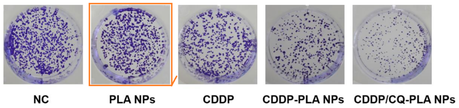
Figure 2 from [9], and Fig 3 from Li & Cao (2020) [5].


And in the darkness measure them
Has everyone had enough of that single ruler? Me neither!

SOURCES
[1]. "Exosomal miRNA-34 from cancer-associated fibroblasts inhibits growth and invasion of gastric cancer cells in vitro and in vivo". Liang Shi, Zhenyong Wang, Xiuchao Geng, Yuhao Zhang, Ziqing Xue (2020) [PubPeer].
[2]. "Pluronic P123 modified nano micelles loaded with doxorubicin enhanced tumor-suppressing effect on drug-resistant breast cancer cells". Zhang et al (2020) [PubPeer].
[3]. "Modification of graphene oxide by angiopep-2 enhances anti-glioma efficiency of the nanoscaled delivery system for doxorubicin". Zhao, Yin & Zhang (2020) [PubPeer].
[4]. "Co-exposure to multi-walled carbon nanotube and lead ions aggravates hepatotoxicity of nonalcoholic fatty liver via inhibiting AMPK/PPARγ pathway". Liu et al (2020) [PubPeer].
[5]. "Pre-incubation with human umbilical cord derived mesenchymal stem cells-exosomes prevents cisplatin-induced renal tubular epithelial cell injury". Li & Cao (2020) [PubPeer].
[6]. "Glypican-1-targeted and gemcitabine-loaded liposomes enhance tumor-suppressing effect on pancreatic cancer". Mu et al (2020) [PubPeer].
[7]. "Transcriptional regulation of miR-483-3p mediated by IL-6/STAT3 axis promoted epithelial-mesenchymal transition and tumor stemness in glioma". Xu, Chi & Xu (2020) [PubPeer].
[8]. "The improved anticancer effects of Bortezomib-loaded hollow mesoporous silica nanospheres on lymphoma development". Li et al (2021) [PubPeer].
[9]. "Multiwalled carbon nanotubes inhibit cell migration and invasion by destroying actin cytoskeleton via mitochondrial dysfunction in ovarian cancer cells". Ping Zhang, Jiangyan Teng, Lijuan Wang (2020) [PubPeer].
[10]. "Antitumor effect of poly lactic acid nanoparticles loaded with cisplatin and chloroquine on the oral squamous cell carcinoma". Li et al (2021) [PubPeer].
[11]. "Multiwalled carbon nanotubes co-delivering sorafenib and epidermal growth factor receptor siRNA enhanced tumor-suppressing effect on liver cancer". Zhili Wen, Yuliang Feng, Youwen Hu, Lingyan Lian, Hongyan Huang, Li Guo, Shanwen Chen, Qian Yang, Moran Zhang, Lijun Wan, Kedong Xu, Degejirifu, Xiaohua Yan (2021) [PubPeer].
[A]. "Effect of miR-21 on apoptosis in hepatoblastoma cell through activating ASPP2/p38 signaling pathway in vitro and in vivo". Liu et al (2019), Artificial Cells, Nanomedicine, and Biotechnology [PubPeer].
[B]. "miR-302e Suppresses Glioma Progression by Targeting VEGFA". Xie et al (2020), Cancer Management and Research [PubPeer].
[C]. "Silencing lncRNA TUG1 Alleviates LPS-Induced Mouse Hepatocyte Inflammation by Targeting miR-140/TNF". Liu et al (2021), Frontiers in Cell and Developmental Biology [PubPeer].
No comments:
Post a Comment