
This post was earlier cross-posted at Leonid Schneider's site, hence the nonfrivolity and Explaining Voice. The version there is improved by Leonid's editing and frame-story.
Two researchers at the Indian Institute of Technology / Indian School of Mines have been in the news lately for nanotechnology retractions, as reported here and here by Prasad Ravinranath. We also refer you to Andy Extance (another science journalist with an interest in research integrity who covered the Sharma / Madhuri imbroglio for Chemistry World) in return for him crediting "anonymous @PubPeer commenters and bloggers like @SmutClyde and @schneiderleonid".
Much of the critical activity occurred at the invaluable PubPeer archive*. The full number of papers under scrutiny is not immediately obvious, due to a limitation of the site's search function. Perhaps it never occurred to its designers that a single author could publish more than 40 papers that would later fall under the shadow of "Post-publication peer review". Alternatively, they were worried that researchers would compete to hold the record for the number of contentious publications. Either way, the search function maxes out and only lists the first 40 hits that match an author's or authors' names... it is an unacceptable restriction and I demand my money back.
Anyway, Drs Sharma and Madhuri felt unfairly singled out by all the attention from image-integrity vigilantes and self-appointed blackboard monitors; they did not welcome the evidence that people were reading their papers. Allegations of extortion ensued, and threats of law-suits, and complaints to the Cyber Police.

As proof of the unfairness, Madhuri and Sharma have adduced a number of arguments, for instance citing the number of Ph.D students they have mentored (their collaborators and co-authors on the papers now under scrutiny). Indeed, this is is a useful reminder that in science we were all students once, [pompous voice] just links in the inter-generational chain of cultural transmission, custodians of skills and values that we learn and pass on [end pompous voice]. Sometimes the transmission goes the opposite way and the guru can learn from the śiṣya.
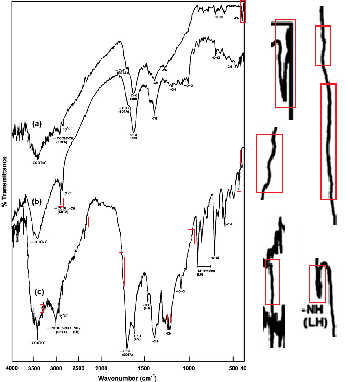

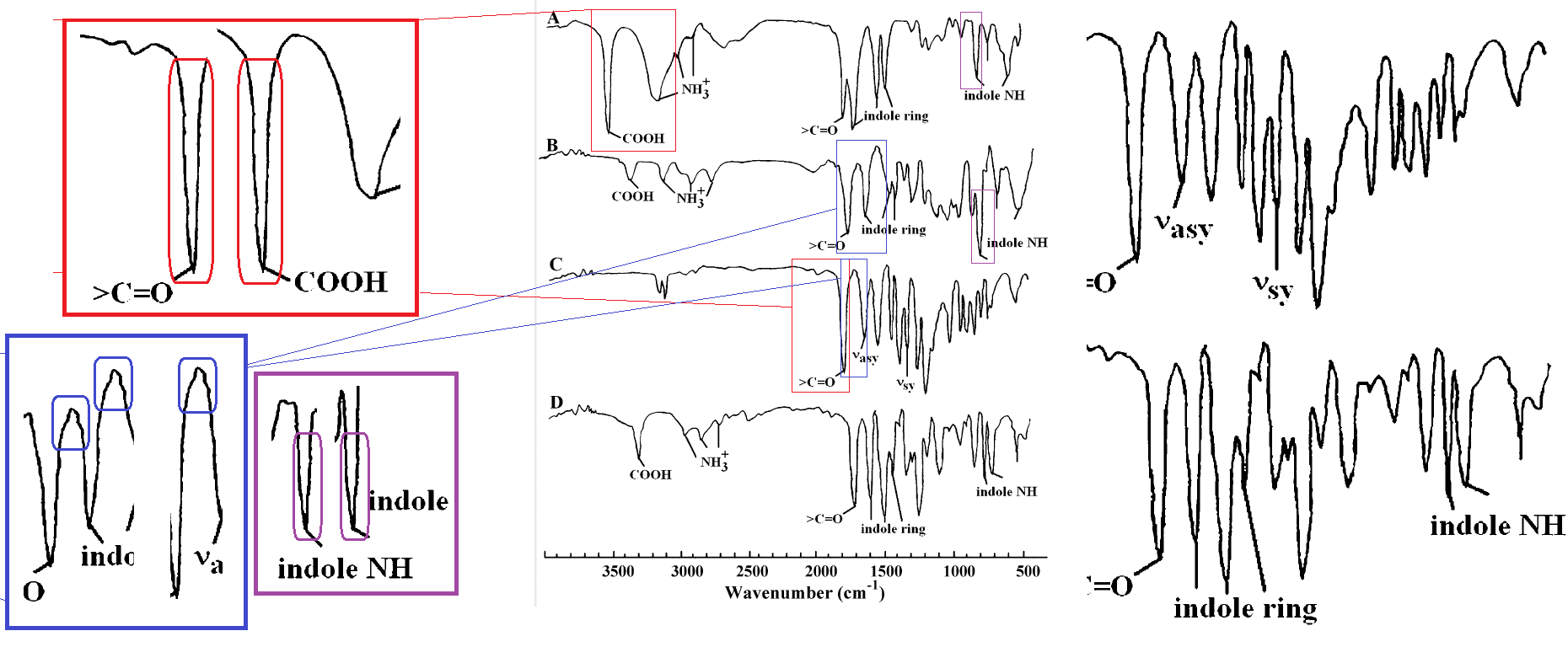
The detector is prepared by coating its surface with what may be a precision polymer, or carbon nanotubes, or quantum dots, or any other combination of nanotech Worship Words. Crucially, the targetted molecule is one of the ingredients during synthesis of the coating, so that the other ingredients wrap around it and form chemical bonds; then the target is washed or stripped away, leaving "negative mold" cavities in the coating, complete with unfulfilled bonds... ensuring that if any target molecules appear in the environs, they will gravitate straight for those cavities and snap back into place with an audible 'click', measurably altering the detector's electrical or physical properties. In many of these publications, the detectors are dual-function ("simultaneous electrochemical determination of norepinephrine and uric acid", or "simultaneous analysis of cerium and gadolinium ions", or "simultaneous analysis of glyphosate and glufosinate"), in the manner of novelty kitchen appliances.
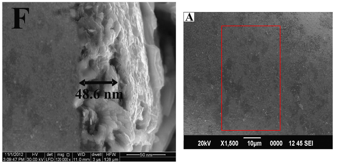
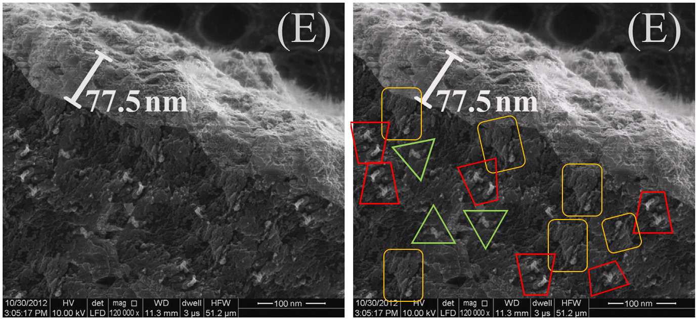
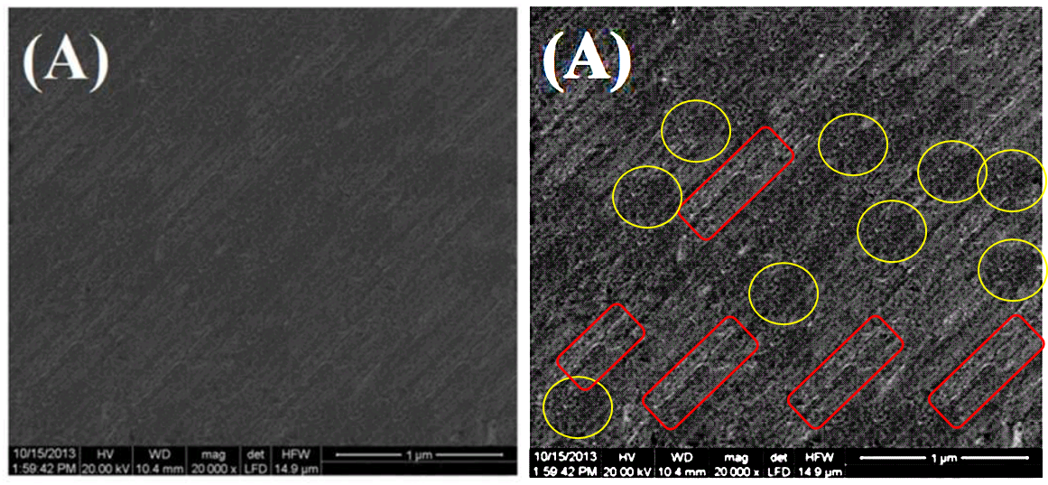
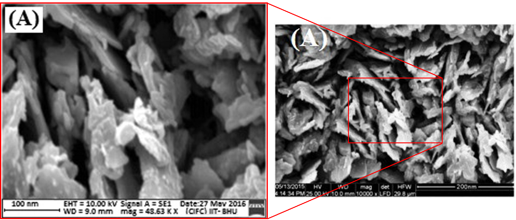
So Prasad's papers are often illustrated with side-views of electrodes, to highlight their magic coatings. This side-view shows either a "OMNiDID/PGE showing film thickness" (according to Fig 1(C) of Prasad et al., 2017), or "MIP- AuNPs films at PGE surfaces" (according to Fig 2(E) of Prasad, Jaiwal & Singh, 2017).

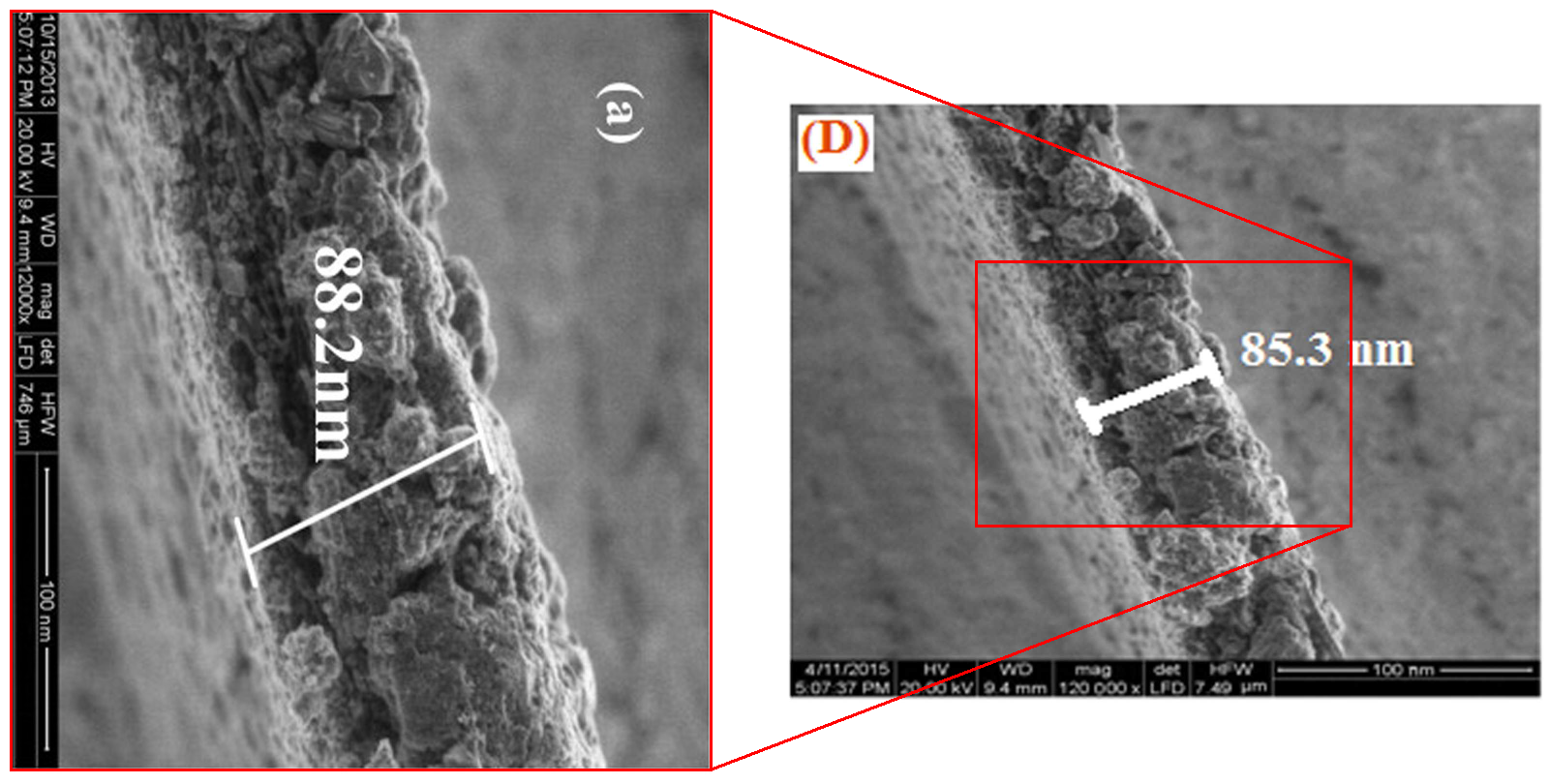
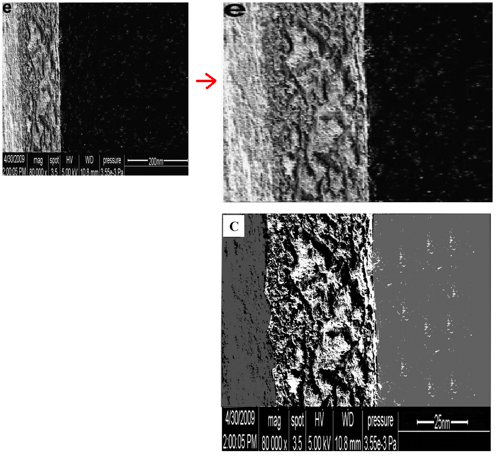
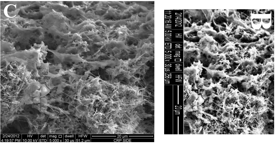
This imposing wall is the edge of either a "MIP-composite modified sandpaper electrode" (Fig. 3C(a) of Prasad & Singh, 2015), or a "C60 monoadduct-MIP" (Fig. 5(D) of Prasad, Kumar & Singh, 2016). We encourage recycling.
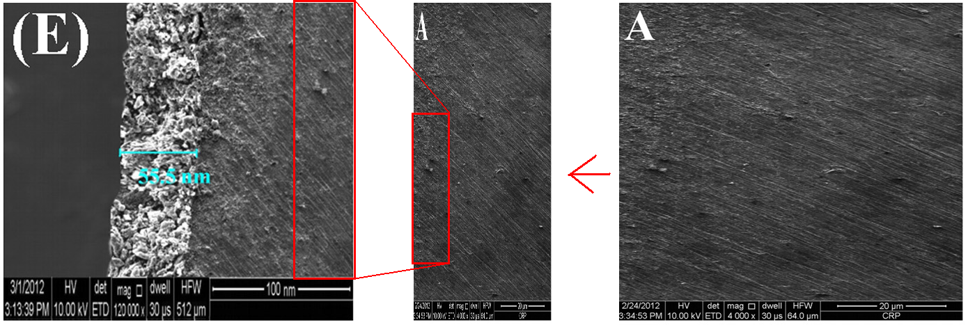
Despite variations in the scale bar, "Fig. 3. SEM images of ... (D) side image of MIP-adduct modified SPCEs" in Kumar & Prasad (2012) is recognisable as "Fig. 2. ... side view of coated layer over PGE, electrode 1 (F)" in Prasad et al. (2013). With a different substrate, it became "Fig. 2. ... (F) side view of coated layer over PGE" in Prasad et al. (2013b).

The background void at the left appears for a fourth time in Prasad, Prasad & Tiwari (2013), as "Fig. 1. SEM images: ... (E) side view of MIP modified MWCNTs–COOH/PGE", with a substrate that was sourced from elsewhere.
"Fig. 1. ...SEM images:... (F) sideview of MIP-modified GNPs-PGE" from Prasad, Jauhari & Tiwari (2014) takes its substrate from Fig. 1, "SEM images of (A) bare ... CIP-modified PGE," of Prasad et al. (2011).
A "MIP-T4 adduct modified Ag electrode for thickness measurement" (Fig 3(e) of Prasad et al. 2010) became a "MIP-AA adduct modified GE (side imaging)" (Fig 2(c) of Prasad et al. 2011). In the course of the transformation, the image lost quality but acquired a grey overlay, while the tastefully cloned 'substrate' at its right was replaced with ugly wallpaper.
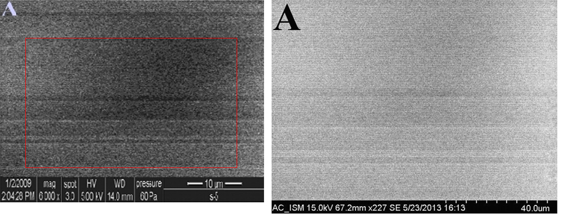
I am not convinced that this "side view of sol–gel–DIIP/PGE" (Fig. 1D from Prasad, Jauhari & Verma, 2014) ever existed outside a Photoshop window...

... or this "side view of MWCNTs/MIP"-Carbon Ceramic Electrode (from Fig. 2(E), Prasad, Jauhari & Tiwari, 2013).
Going back to the molecular-imprinting paradigm: it has an intuitive appeal, though like all the best cargo cults, it is best to avoid inquiring too deeply into the working details. It appeals enough to keep the research grants flowing. Conceivably some actual experiments were conducted, hidden behind the photoshops, and the impostures, and the photocopied hand-drawn spectra and voltagrammetric ziggurats, and the forged time-stamps, and the shifting identifications of electron microscopy.
The photoshop component of this decade-long body of work is variable in quality. It may be that the co-authors / students are expected to contribute a composite image as a kind of journeyman's piece, to demonstrate their mastery of the image-manipulation software that seems to be central to the BHU Analytical Chemistry curriculum. Then of course they take those hard-earned skills (and their training in the values of science) when they graduate and move on to academia elsewhere.
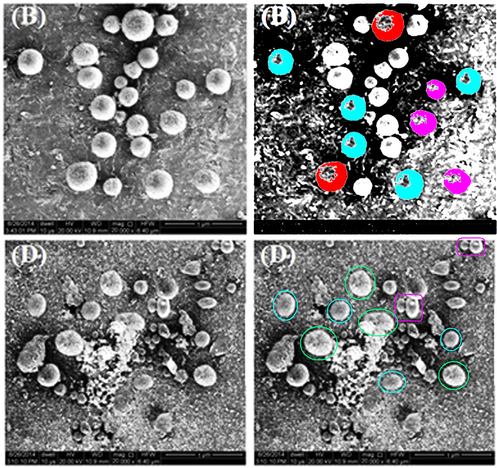
So here is Figure 3(B), a scanning-electron-microphotograph of "anodised silver", from Prasad and Kumar (2014):
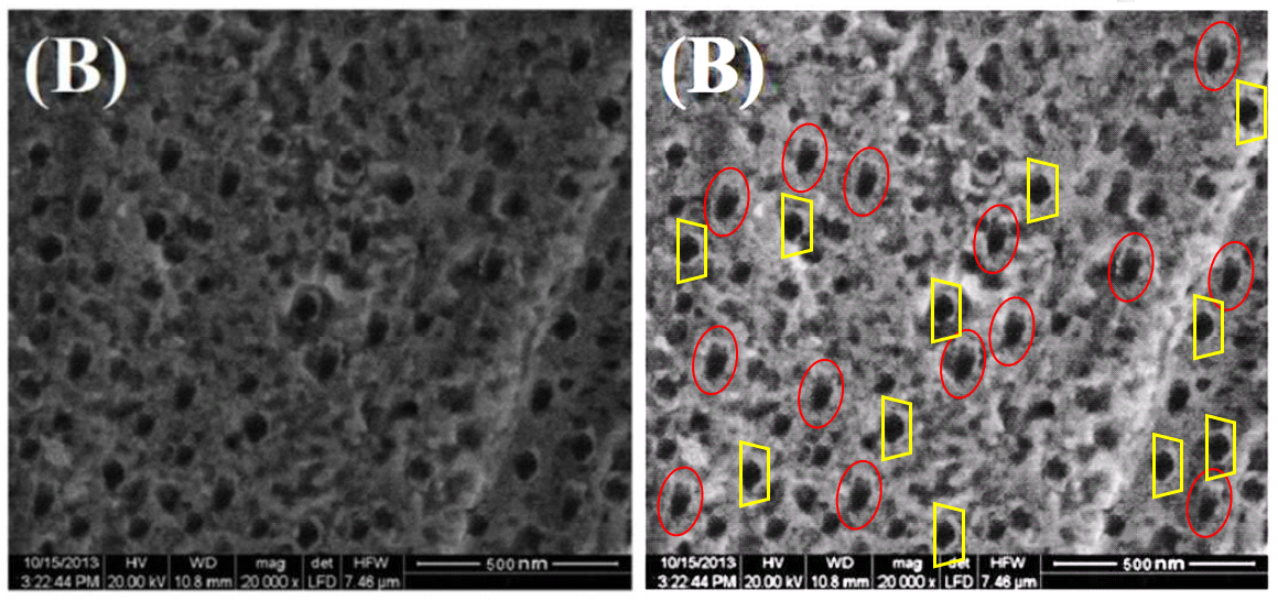
I like the idea that "some of the holes are very small Sasquatch prints, and the round ones were left by a hunter's shotgun". From the same paper, 3(A), "bare silver surface". The patchwork assembly of repeated motifs all seems very labour-intensive and it might have been easier to find a piece of bare silver to put in the SEM.
Sometimes the repetition is so dominant, one could be reading a wallpaper catalog, not Material Science & Engineering. "Was the quintuplicated baboon’s face in (c), as marked up with red in Hoya’s depiction, originally inspired by this example from a tattoo design web site?" wondered C. Amazonicum.

Figure 3(c) and (d), Patel et al., 2009.
"Perhaps there is a desperate EM technician trapped somewhere in the basement?"
Figure 2(A), Prasad & Pandey, 2012
Elsewhere, the original image has been so stirred around by the 'Shop distortion tools as to elude recognition. Assuming that a non-virtual starting-point did exist, though I am open to the possibility that the source was a neural network that someone trained on the depraved perspectives of Matta's ambiguous Surrealist spaces, and H. R. Giger's gauzy airbrushed 'Landscape' series.

Figure 5, Prasad et al., 2016
At their best, these creations become nebulous dream spaces in which foreground and background blur, or vertiginous abysses, reminiscent of Dorothea Tanning.

Figure 3(a) and (b), Patel et al., 2010

We need to move on, but I cannot resist showing Fig. 3(D) – "MID nano-fiber at 75,000×" (from "A dual-template biomimetic molecularly imprinted dendrimer-based piezoelectric sensor for ultratrace analysis of organochlorine pesticides": Prasad & Jauhari, 2015) – for I am in the thrall of the meshes of its intricate symmetry. I think it was modeled on the Veve of Ogoun.

As for Fig. 3(C) ("MID-templates adduct nano-fiber at 75,000× magnification"), it seems to have started out as 3(F) of Prasad et al. (2010) – "MIP (after template extraction)-modified Ag electrode". But elongated, and half-obscured by exuberant use of the 'Shop Clone Stamp.**

Figure 3(A) from Prasad et al. (2016b) is as if the loosely-stacked books on my shelves sprouted eyes and stared at me reproachfully. Or like air-dried codfish heaped up in a Norwegian farm-house.
In this case, a source for the composition can be identified, for the central portion later appeared as Figure 2(A) of Prasad et al. (2017).
This is a convenient segue leading to another part of this body of pictorial invention, in which parts of the confections are used elsewhere in different contexts. That is to say, there is no internal repetition, so it is easier to forgive the journal editors and peer-reviewers for blithely ignoring their dubious nature.
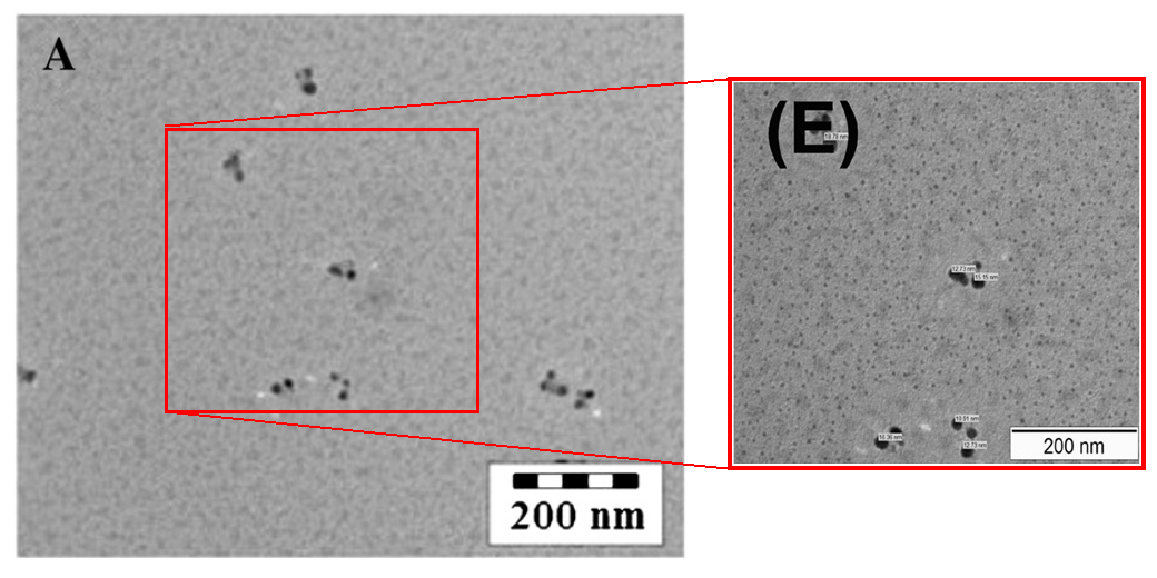
At one extreme are the simple rotations, and zooms, and unadorned re-use as representations of a different nanomaterial. There are so many cases of 'enlargement' as an aid to recycling, I don't know which ones to leave out. Readers will just have to explore the PubPeer archives for themselves. This transmission-electron-microphotograph shows either TNPs, or Quantum Dots.
"Fig. 1. SEM images: ... (E) MIP," from Prasad et al. (2014), or "Fig. 2. SEM images: (A) MIP-adduct" from Prasad et al. (2017)?
SEM images of "MIP modified PGEs" (Fig 5(E) of Prasad et al., 2016), or "NIP–carbon composite fibers at 10,000 x" magnification (Fig 2(F) of Prasad et al., 2010)?
At the moment my favourite image manipulation is Figure 3 from Prasad et al. (2010), in which (A) and (B) ("TEM images of crude and MIP modified MWCNTs) are simply the zoomed-in centres of photo negatives of (E) and (D) respectively ("SEM images of MWCNTs-NIP. ... MWCNTs-MIP").***

Moving right along, the "MIP-adduct" (Fig 3(C) from Kumar & Prasad 2012) is a 90° rotation of "MIMSPE fiber: MIP (after template extraction)" (Fig. 1(B) from Prasad et al. 2014). Both also have a sense of vibrant, interrupted movement that somehow brings "Neptune's Horses" to mind... if a fondness for late-Symbolist painting is wrong then I don't want to be right.
.jpg)

A mystery lies behind the "bare pencil-graphite electrode" that appeared with a 2009 electron-microscopy time-stamp as Fig. 3(C) of Prasad et al. (2010) and as 2(A) of Prasad et al. (2010b), for the stated magnifications are vastly different (60,000 x and 8,000 x respectively). When the same image featured as Figure S3A in Prasad et al. (2011), the date-stamp had changed to 2010. Co-author Madhuri must have taken the image with her when she graduated, for a zoomed-in version appeared for a fourth time in Roy et al. (2014), re-purposed as a platinum electrode.
But these digital, virtual fabrications of results are not to everyone's taste, and it is good to be able to say that old-school analog methods are not neglected. Figure 3 of Prasad et al. (2010) is nominally the output of an IR spectrograph, but "the wobbly palsied lines that compose these overhanging, toppling stalagmites" were more likely hand-drawn with a dried-out felt-pen, photocopied and reassembled... keeping it real, as the kids say.

If you prefer vinyl records and valve-amplifier stereros to MP3 files, you are sure to like Figure 4 of Prasad and Lakshmi (2005). It is presented as a series of voltage / current functions - "Differential pulse cathodic stripping voltammograms with MIP-modified hanging mercury drop electrode" - plotted on a X-Y chart recorder, but I believe it to be some kind of hand-drawn ziggurat construction diagram.

Often photocopies of a crennelation sketch are stacked up, to show how well the voltagrammetry replicates. At left is Fig. 5(C) from Prasad, Srivastava &Tiwari (2013): "run c, multiple runs show reproducibility of the measurement".
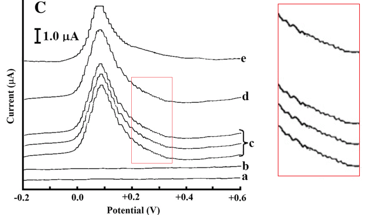
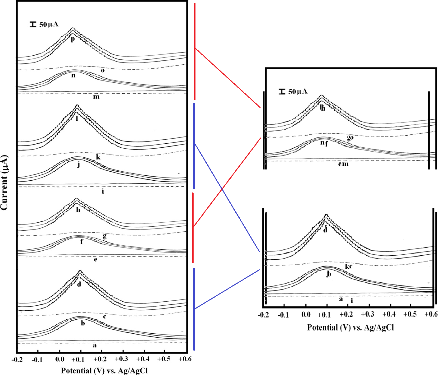
At right, Fig. 2 from Prasad, Srivastava & Tiwari (2013b).
* Obligatory caveat: There is no rule that commentary at PubPeer has to be negative; anyone can use the platform to praise a paper and bring it to wider attention, or for that matter, to criticise it on spurious or malicious grounds - thus the existence of a PubPeer thread for a publication is not damning in itself.
** 3(F) also manifests as a SEM image of "MIP modified QCM sensor", in Figure 2(a) of Madhuri et al (2011).
*** 3(D) reappeared as a "NIP-MIMSPE fiber" (as Figure 1(C) of Prasad et al. 2014); and yet again as a "MIP modified PGE surface" (as Fig. 1(a) in Kumar et al. 2011).
3 comments:
I love these, smut, yer doin' a fine job. Can you really make money with just Photoshop & a bunch of scientific bafflegab? I got into the wrong line of work, clearly...
Evidently you can sustain a career as Professor and Head of Department, gubblement advisor, and recipient of prizes and research grants.
The scientific Bafflegab is a prerequisite. The Photoshopping is just in case someone timidly opines that the Bafflegab means nothing.
Post a Comment